Palm oil may up risk of obesity, diabetes, cardiovascular diseases, cancer | 棕櫚油可能會增加肥胖、糖尿病、心血管疾病、癌症的風險

中文版谷歌中文翻譯(90% 準確率) | English translation
Buy/Sell Your Domains Here。在這裡購買/出售您的域名
Contact Dr. Lu for information about cancer treatments。聯繫盧博士,獲取有關癌症治療資訊。

Editor’s note:  This is an excellent review on palm oil and its health impact.  Everyone who likes processed foods should read this article.  In a nutshell, palm oil is not a healthy oil.  (Any added cooking oil can pose a health risk, but palm oil is worse than others such as sunflower oil, olive oil and canola oil).  Palm oil is used because its unique high-melting point which is important for dry processed foods)

編者註:這是一篇關於棕櫚油及其健康影響的優秀評論。 每個喜歡加工食品的人都應該閱讀這篇文章。 簡而言之,棕櫚油不是一種健康的油。 (任何添加的食用油都會造成健康風險,但棕櫚油比葵花油、橄欖油和菜籽油等其他油更糟糕)。 使用棕櫚油是因為其獨特的高熔點對乾加工食品很重要)

Biological and Nutritional Properties of Palm Oil and Palmitic Acid: Effects on Health

1
Dipartimento di Scienze Motorie e del Benessere, Università di Napoli “Parthenope”, via Medina 40, Napoli 80133, Italy
2
CEINGE Biotecnologie Avanzate s.c.a r.l., via G. Salvatore 486, Napoli 80145, Italy
3
Dipartimento di Scienze e Tecnologie Ambientali Biologiche Farmaceutiche, Seconda Università degli Studi di Napoli, via G. Vivaldi 42, Caserta 81100, Italy
*
Author to whom correspondence should be addressed.
These authors contributed equally to this work.
Academic Editor: Maurizio Battino
Molecules 201520(9), 17339-17361; https://doi.org/10.3390/molecules200917339
Received: 31 July 2015 / Revised: 2 September 2015 / Accepted: 9 September 2015 / Published: 18 September 2015

Abstract

A growing body of evidence highlights the close association between nutrition and human health. Fat is an essential macronutrient, and vegetable oils, such as palm oil, are widely used in the food industry and highly represented in the human diet. Palmitic acid, a saturated fatty acid, is the principal constituent of refined palm oil. In the last few decades, controversial studies have reported potential unhealthy effects of palm oil due to the high palmitic acid content. In this review we provide a concise and comprehensive update on the functional role of palm oil and palmitic acid in the development of obesity, type 2 diabetes mellitus, cardiovascular diseases and cancer. The atherogenic potential of palmitic acid and its stereospecific position in triacylglycerols are also discussed.

1. Introduction

Over the last few decades there has been growing public concern about the significant interplay between health and nutrition. Fat is an essential macronutrient of the human diet and vegetable oils represent a more highly consumed fat. The effects of high-fat diet, mainly in saturated fatty acids (SFA), have been the focus of several dietary guidelines targeting the reduction of cardiovascular diseases (CVD) [1,2], obesity-related diseases and, recently, cancer prevention [3].
The increasing demand for vegetable oils is a worldwide phenomenon and palm oil (PO) contributes significantly to the global supply of edible oils. PO is entirely GMO-free and produces up to 10 times more oil per unit area than other oilseed crops. In 2012 PO accounted for 32% of global fats and oils production and it has overtaken soybean oil as the most important vegetable oil in the world [4].
The palm tree (Elais guineensis) is an ancient tropical plant native to many West African countries, where local populations traditionally use its oil for cooking and other purposes. Large scale palm crops are found across the tropical regions, and Malaysia and Indonesia are the leading producers of PO, accounting for the 86% of global production; other PO producing countries are Nigeria, Thailand, Colombia, Papa Guinea, Cote d’Ivoire, India and Brazil.
Two different types of oil are extracted from palm fruit: palm kernel oil (PKO), from the seeds and PO, from the mesocarp. The edible oil contained in the mesocarp of palm fruit can be extracted using different methods, with the most common known as wet or dry processes [4].
Crude palm oil (CPO, known also as red palm oil, RPO), extracted either by wet or dry processes, contains both healthy beneficial compounds, such as triacylglycerols (TAGs), vitamin E, carotenoids, phytosterols, as well as impurities, such as phospholipids, free fatty acids (FFAs), gums, and lipid oxidation products; the latter can be removed by means of refining processes [5]. Hence, the CPO is purified by centrifugation and drying; the dried oil is then cooled and stored in appropriate containers [6].
CPO represents the richest natural source of carotenoids (500–700 ppm), tocopherols and tocotrienols (600–1200 ppm), all contributing to its stability and nutritional properties [4,5,7,8,9]. Their antioxidant properties, exerted mainly against reactive oxygen species (ROS), play a role in aging, in CVD and in cancer prevention [7,10,11]. Furthermore, tocotrienols have been reported to be natural inhibitors of cholesterol synthesis [7].
Despite the good quality of CPO, food manufacturing industries require PO with a bland and light color, making refinery a mandatory step. These steps can be carried out via chemical (alkaline or acid treatment) [12,13] or physical methods (steam refining, inert gas stripping, molecular distillation, membrane refining, etc.) [13,14]. Afterwards, bleaching and deodorizing steps follow, along with other specific treatments depending on the first refining process applied. For example, after alkali treatment, the impurities are removed by centrifugation as gums; volatile products are then removed through stream distillation at higher temperature and reduced pressure. The oil obtained after the refining processes is colorless, bland and stable. On the other hand the alkali treatment produces losses in neutral TAGs and it is a more expensive process with a high impact on the environment. For these reasons, physical refining protocols, based on different volatility of FFAs and TAGs, high-temperature and low-pressure methods, are currently used [13,14], even if these procedures lead to an evident loss of tocopherols and tocotrienols and also produce oxidative damages [13]. Moreover, PO refined by physical treatments shows lower storage stability and require an accurate procedure in order to eliminate phosphorus during the degumming procedure, to avoid the presence of phosphoric acid in the final product that would affect the heat bleaching process [13].
PO with low amounts of free fatty acids (FFAs), low impurity content and good bleaching is considered of high quality and used in the edible oil industry; conversely, low-quality oils are used in non-edible industry such as biofuel, candles, cosmetics and soap production [15]. So-called high-quality PO is constituted of more than 95% neutral triacylglycerols (TAGs, or triglycerides) and less than 0.5% FFAs [13,14].
PO and PKO have different physical and chemical properties depending on their intended applications [4,10]. As shown in Table 1, PKO contains 85% SFAs, mainly lauric and myristic acids, while PO contains 50% SFAs, mostly palmitic acid (PA, 44%) and lower amounts of stearic acid (5%), 40% monounsaturated fatty acids (MUFAs), mostly oleic acid, and 10% polyunsaturated fatty acids (PUFAs), mostly linoleic acids [5,7,16]. PA is the main SFA naturally occurring in animal fats and vegetable oil, as well as the main component of human milk fat [17].
Table 1. Fatty acid composition of palm oil and palm kernel oil.
In the last few decades, PO’s application in food industries has exponentially grown for the texture, the fragrance and the neutral taste it guarantees in the finished products. Its two major fractions are the low-melting liquid fraction (known as palm olein, 65%–75%) and the high-melting solid fraction (known as palm stearin, 30%–35%). The different palm oil fractions are then differently used in food industry: palm olein is used in cooking oil for frying (because of its high smoke point, 230 °C) and margarines; palm stearine in food applications such as shortenings and hydrogenated oils used as butter substitutes in India; double-fractioned palm olein is then used in mayonnaise preparation [4]. PO is generally found in: baked goods, candies, cakes, cheese analogs, chips, chocolate, confectionary fats, cookies, cooking oil, crackers, doughnuts, frozen meals (pancakes, pies, pizza, potatoes), ice cream, industrial frying fats, instant noodles and oatmeals, margarines, microwave popcorn, non-dairy creamers, peanut butter, salad dressings, snacks, soups, supplements/vitamins, vegetable ghee.
Despite some authors suggesting that palm olein, containing oleic acid at 48%, could be a substitute for olive oil in healthy normo-cholesterolaemic human diet [5], debates related to PO’s potential unhealthy effects (mainly due to the high PA content) are ongoing [18,19,20,
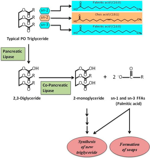
21]. However, PO, like all vegetable oils, is cholesterol-free and FAs are mainly structured as TAGs. TAGs contained in PO are mainly constituted of oleic acid, predominantly located at the SN-2 position, and of PA, mainly located at the SN-1 and SN-3 positions (Figure 1). TAGs contained in animal fat are different in that PA or stearic acid are often found at the SN-2 position [5,22].
Figure 1. A schematic representation showing the hydrolysis of typical PO triglyceride. Palmitic acid, the main SFA in PO, is situated at SN-1 and SN-3 positions of TAGs. The other main FA in PO, oleic acid, is situated at SN-2 position. After digestion, FFAs are involved in the re-synthesis of new triglycerides and/or formation of Ca2+ or Mg2+ soaps. Free fatty acids, FFAs; palm oil, PO; saturated fatty acid, SFA; triglycerides, TAGs.

2. Palm Oil and Palmitic Acid: Role in Obesity and T2DM

Obesity is a metabolic disease characterized by an excess of white adipose tissue (WAT) resulting from an excess of energy stored in adipocytes as TAGs, responsible for adipocyte hypertrophy and hyperplasia [23]. In recent years, obesity has assumed epidemic proportions: about 1.9 billion adults worldwide and 42 million children below 5 years are obese [24]; morbidity and mortality associated with obesity-related diseases, such as type 2 diabetes mellitus (T2DM), CVD, hyperlipidemia and hypertension, have increased exponentially [25].
WAT produces adipokines that are responsible of chronic inflammation processes and are associated with obesity-related metabolic diseases [26]. Adipose tissue from obese mice contains elevated SFA depots that activate Toll-like receptor 4 (TLR4)-mediated inflammatory signaling [27,28,29].
SFAs also stimulate pro-inflammatory mechanisms through reactive oxygen species (ROS) in a TLR-independent manner. ROS regulate the activation of mature interleukin-1β (IL-1β) from its inactive precursor pro-IL-1β [30,31]. As a consequence, IL-1β down-regulates the insulin-signaling in insulin target cells, providing a possible SFA-mediated inflammatory response resulting in insulin resistance [32,33].
More recently, a link was suggested among high-fat diet, inflammation and pro-inflammatory products in plasma originated in the gut by the death of Gram-negative intestinal microbiota, namely lipopolysaccharides (LPSs) or endotoxins [34,35]. LPSs, bound to LPS-binding proteins, trigger inflammation via the TLR4 pathway and induce secretion in the plasma of pro-inflammatory cytokines, such as IL-6 [36]. Moreover, several studies showed that metabolic endotoxemia could be generated by gut LPS absorption during digestion of a high-lipid load [37,38]. A direct effect of PA on increased secretion of IL-6 in 3T3-L1 adipocytes, compared to myristic, linoleic or α-linolenic acids, was reported by Laugerette and co-authors [39]; they also demonstrated the cooperative effect of PA and LPS co-treatment in these cells, confirming the role of LPS in modulating inflammation through different dietary fat contents. Similarly, the same authors found that PO-enriched diet induced higher amounts of inflammatory markers in plasma (IL-6) and in WAT (IL-1β, TLR4 and CD14) compared to other fat-based diets in mice [39].
There is growing evidence linking obesity to changes in gut microbiota [40,41]. PO-rich diet determines weight-gain and hepatic lipid accumulation compared to unsaturated fat diet (olive or safflower oil) in C57BL/6J mice, together with a reduced microbial diversity in the intestine [
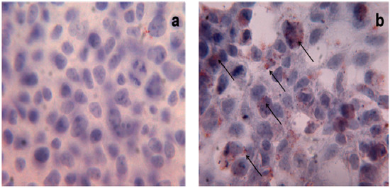
42]; these observations support the hypothesis that an excessive consumption of PO in the diet triggers changes in gut microbiota components and determines lipid accumulation. Triglyceride-droplet accumulation mediated by high doses of PA (500 μM and 1 mM) has been also described in human HEK293 cells (Figure 2) [43]; interestingly, the authors highlighted the role of uncoupling protein-3 (UCP3) in the metabolism of long-chain fatty acids, such as PA, in these cells.
Figure 2. Triglyceride storage in HEK293 cells. Oil Red O staining of untreated (a) and treated (b) HEK293 cells with PA 1 mM. Red points indicate triglyceride depots; ×40 magnification.
The effects of PO, and of its main constituent PA, have also been investigated on other organs and tissues, such as the central nervous system (CNS).
The effects of intra-cerebroventricular (icv) injection of PA on hypothalamic leptin signaling, inflammatory marker secretion and hepatic energy metabolism were recently analysed in C57BL/6J male mice [44]. The authors reported that high-doses of PA induce pro-inflammatory responses and leptin resistance, similarly to obesogenic-diets; however, the key role of PA in the hypothalamus in response to high-fat diets has not been fully characterized yet.
Another interesting aspect is the relation between diet, with different content in lipids consumed during pregnancy and lactation, and development of obesity in adult life. The fetal nutritional environment is involved in obesity and/or obesity-related diseases in later life [45,46]. Several studies carried out on animal models indicated that, during lactation, the consumption of normolipidemic diets, rich in SFAs derived from PO and/or in partially hydrogenated fats (PHF) induces fat retention in the young offspring [47]. In fact, maternal intake of PO and inter-esterified fat (IF) present in processed foods predisposes the offspring to the development of obesity in adult life [48].
In obesity, an altered production of pro-inflammatory cytokines, such as TNF-α, IL-6, leptin and adiponectin, affects both secretion and efficiency of insulin action, hallmarks of T2DM, along with progressive failure of β cells [49,50,51]. In fact, obesity and T2DM are strongly interconnected and their overlap leads to predisposition to a large number of metabolic diseases, including metabolic syndrome [52,53].
Oxidative stress and ROS overproduction are significantly increased in T2DM, promoting endothelial dysfunction, a condition more prone to the pathogenesis of hypertension and CVD in diabetic patients. Recently, T2DM was also linked to immune system activation and inflammation [54].
The role of PO consumption in T2DM is controversial in that, it is difficult to exactly distinguish between the effects of PO and/or other fat components of the diet in T2DM risk. Some evidence indicates that diet supplemented with PO impairs glucose tolerance in mice [55]; this finding may be ascribed to the reduction in insulin-sensitivity induced by PO-enriched diets and to the corresponding increase of serum TAGs; in fact, elevated serum TAGs are associated with insulin-resistance [56]. Furthermore, several studies, involving animals and humans, have shown that fatty acid composition of diet affects muscle membrane phospholipid dynamics (such as fluidity) and the ligand/receptor recognition process (such as insulin/insulin receptor) [57]. On rat epididymal fat cells, PO treatment resulted in a lower rate of insulin-stimulated glucose uptake due to reduced insulin binding to the cells compared to sunflower oil treatment [58].
Studies conducted on humans yielded conflicting results: four weeks of PO treatment in 30 patients affected by T2DM had no significant effects on plasma glucose concentration [59]; on the contrary, Rosqvist and co-authors demonstrated in 39 T2DM patients that an excessive consumption of SFAs (among which PA) favors both liver and visceral fat accumulation with detrimental role in T2DM development and progression [60]. Also the largest study on T2DM patients, the EPIC-InterAct case-cohort study, was not indicative. In fact, the study, including 12.403 Caucasian people with incident T2DM, reported that different SFAs (myristic, palmitic, and stearic acid) have different effects on T2DM incidence. Interestingly, the authors underlined that PA can also be synthesized endogenously through de-novo lipogenesis, a metabolic pathway stimulated by increased intake of carbohydrates and alcohol; these evidences introduce a further complexity level in this puzzle making very difficult to distinguish between endogeneous and exogeneous PA effects on T2DM onset risk [61]. Furthermore, in vitro studies demonstrated that PA directly impairs insulin signaling in cultured rat hepatocytes and myotubes as well as pancreatic cells vitality and insulin secretion [62]. Three main mechanisms have been suggested: (1) reduction of viability and induction of apoptosis through ER stress. In rat insulinoma cells, PA provokes toxicity through lipotoxic stress in ER; this mechanism, exerted by different SFAs, has been described in various cell types [50,63,64,65]. In addition, SFAs can directly interact with ER membrane bilayer disturbing ER morphology and function [57]; (2) inhibition of insulin-stimulated phosphorylation event. PA markedly impairs phosphorylation and, therefore, activation of insulin receptor, insulin receptor substrate-1, and Akt in several cell types, contributing to the onset of insulin-resistance [66]; (3) ubiquitination of the key insulin-signaling molecules. PA facilitates the ubiquitination and, hence, elicits proteasome degradation of insulin receptor, insulin receptor substrate-1, and Akt [67].

3. Palm Oil and Palmitic Acid: Role in CVD

CVD represent the principal cause of death worldwide. Main serum/plasma CVD-related biomarkers are TC, LDL-C, high-density lipoprotein cholesterol (HDL-C), TAGs and very low-density lipoprotein cholesterol (VLDL-C). Also apolipoprotein A-I (apo A-I) and B (apo B) reflect variations in HDL-C and LDL-C, respectively [68,69]. In particular, low apo A-I and high apo B levels are associated with increased CVD risk and the apoB/apo A-I ratio is considered a good CVD predictor factor [70].
The link between PO consumption and CVD risk refers to PA content in PO, which accounts for 44% of total fat (Table 1). Since 1950 Toshima’s group demonstrated that high contents of SFAs in diets are powerful predictors of heart diseases, the authors provided a specific equation to predict serum cholesterol levels starting from SFA intake [71,72,73,74]. Successively, the “lipid theory” stated that an excessive SFA consumption is responsible for hypercholesterolemia, a condition that causes predisposition to a higher CVD risk: an increase of 20 mg/mL in total serum cholesterol determines a 12% higher CVD risk [75]. At the same time, Mensink and co-authors [76,77] demonstrated that the most favorable lipid profile is obtained when MUFAs or PUFAs replace SFAs.
In a multi-country analysis Chen and co-authors [78] evaluated the effects of PO consumption on CVD mortality-risk due to ischemic heart disease (IHD) and stroke. Twenty-three countries were analyzed and divided into historically high-income countries (HICs) and developing countries (DCs). The authors reported that every additional kg of PO consumed per-capita annually determined higher IHD mortality rate in DCs respect to HICs. The same trend was also observed in stroke deaths, but in this case, data were not statistically significant, suggesting that serum LDL-C is not strictly linked to hypertension.
Nevertheless, it is now emerging that both favorable and unfavorable changes in CVD biomarkers are observed when PO constitutes the main dietary SFA intake [19,20]. In studies where PO was compared to soybean oil (a vegetable oil with more PUFAs and less SFAs) [19,79], to olive oil (rich in oleic acid, a MUFA) [19,80], to sunflower oil (rich in oleic acid and PUFA) [19,80] and to canola oil (rich in MUFAs) [19,81], no substantial differences in the lipid serum profile were observed. In some cases, changes in cholesterol fractions, not affecting TC/HDL-C or LDL-C/HDL-C ratios, were registered. Furthermore, other studies failed to unequivocally demonstrate the association of PA consumption and CVD risk increase, particularly in normocholesterolemic subjects assuming the recommended PUFA intake [82,83]. Finally, SFA intake, associated with PO consumption, did not significantly affect the apoB/apoA-I ratio [84,85,86].
In addition, it is well known that the number of carbons in the FA chain, the degree of saturation and the stereospecific positioning of FAs in TAG structures highly influence FA absorption, thus affecting their metabolism and their role in CVD [10,87,88]. In fact, CVD biomarkers (TC, LDL-C, HDL-C and VLDL-C) worsen when main dietary SFAs are characterized by shorter carbon chains: lauric (C12:0) and myristic acids (C14:0) increase all the cholesterol fractions (favorable and unfavorable) more than PA (C16:0); this latter increases all the cholesterol fractions (favorable and unfavorable) more than stearic acid (C18:0) [89,90]. Furthermore, the atherogenicity of a specific TAG is associated to the degree of saturation of FA located at SN-2 position [91,92,93,94,95]: in fact, it has been shown that higher percentages of PA at SN-2 position are related to the most atherogenic profiles. Accordingly, Kritchevsky and co-authors reported that lard is highly atherogenic because almost all PA (21.4%) is at SN-2 position in the TAGs. Interestingly, when a randomization process is applied to modify the TAG structure, the presence of PA at SN-2 position is reduced by about 67% and, consequently, the atherogenic potential decreases to the half [93]. Likely, tallow has a similar content in PA (24.8%) with a smaller fraction (3.8%) being at SN-2 position; following randomization, the amount of PA at SN-2 position increases by about 124%, and the atherogenic potential raises [93]. Comparing different animal fats and vegetable oils, the percentage of PA in SN-2 position of corresponding TAGs is 66% in animal butter, 58% in human milk, 34% in bovine milk, 4.4% in PO and 0.3% in olive oil [88]. In fact, as shown in Figure 1, the typical TAGs of PO are symmetrical with PA bearing the SN-1 and the SN-3 positions and oleic acid the SN-2 position. Long chain SFAs impair absorption rates from the intestinal lumen when they occupy SN-1/SN-3 position of glycerol backbone; following lipase action, they are converted in FFAs and tend to form insoluble soaps, excreted in faeces [96], instead of being transformed into bile salt micelles or vesicles [87]. These evidences are associated with a reduced atherogenicity and support the concept that PO carries a low CVD risk within balanced diets [10].
The atherogenic effects of PO were also compared to CPO and to reconstituted CPO (PO added with carotenoids, tocotrienols and tocopherols) [95] in rabbits fed with semipurified diets containing 0.1% cholesterol and 13% test oil (PO, CPO, reconstituted CPO) for 90 days. At the end of the treatments, serum cholesterol levels were comparable among rabbit groups, whereas reconstituted CPO determined a higher increase of TAGs. Authors hypothesized that the fine structure of native CPO is different from the reconstituted CPO, and that the absorption of carotenoids and vitamin E in native CPO could be different, leading to the idea that natural products cannot be easily reproduced by simply adding and mixing specific components.

4. Palm Oil and Palmitic Acid: Role in Cancer

There is now considerable evidence that, in addition to the known risk factors, dietary fat intake plays an important role in determining cancer risk [97,98,99]. For decades, epidemiological studies indicated a positive association between total fat intake and risk of breast, colorectal and prostate cancers. Starting from this evidence, it has been postulated that a high-fat diet (>25% FAT) induces a major risk of cancer development compared to low-fat diet (<20% FAT). The dietary fat hypothesis is supported by several meta-analysis studies obtained from data collected up to nowadays [100,101,102]. Retrospective case-control investigations also report a relationship between increased fat intake and higher cancer risk with conflicting results.
In a recent prospective study in postmenopausal women, the authors found positive associations between total MUFA intake or PA and stearic acid intake and breast cancer incidence [97]. In agreement with these data, a previous meta-analysis on prospective studies revealed that high levels of PA were associated with an 89% increased risk of postmenopausal breast cancer [100]. Although this study supports the hypothesis of breast cancer risk associated with fat consumption, most of the prospective studies failed to confirm this hypothesis, reporting inverse associations [103,104] or no associations [105].
To our knowledge, only a few studies reported significant increased risks of breast cancer associated with MUFA consumption [106].
Interestingly, a national prospective case-control in Scotland showed that PA, together with total MUFAs and others SFAs, were positively associated with colorectal cancer risk in a dose-dependent manner; on the other hand, these effects were abolished after adjusting for potential confounding factors such as family history of cancer, total energy and/or fiber intake, drugs, smoking, body mass index, and physical activity [107].
As for prostatic cancer, epidemiological studies lack to find a consistent association between FA intake and cancer risk. However, a case control analysis conducted on plasma FAs revealed a positive association between PA consumption and risk of localized and low-grade prostate cancer [108]. Furthermore, a prospective cohort study in Japan showed that PA increased the risk of prostate cancer in a dose-dependent manner [109], whereas higher intakes of dietary MUFA as well as moderate concentrations of plasma PA were inversely related to prostate cancer [99].
The conflicting results of these studies may be due to (i) sample size that, in same case, was not large enough to observe statistical significant positive associations; (ii) use of different biomarkers of FA intake (blood or adipose tissue) with different time-frame exposure: in fact, FA profile of adipose tissue reflects the relative intake over 2 years, whereas FA composition of blood indicates recent intake; (iii) the heterogeneity of study populations that display different characteristics (i.e., age, hormonal status, body mass index, physical activity), additional cancer risk factors (obesity, weight loss, smoking, drugs and alcohol) and other pathological conditions (presence or not of cancer) affecting lipid metabolism and, as a consequence, FA composition.
Different effects of SFAs, such as PA, and MUFAs, have been reported on cell proliferation and tumor development also in in vitro and animal studies [110,111]. The conflicting results may be due to a combined administration in animals of different FAs. However, such results raised new questions about the key role played by the type of FAs (saturated, monounsaturated, and polyunsaturated) besides to their amounts.
It has been reported that PUFAs of the linoleic group (n-6 PUFAs) stimulate breast, colorectal and prostate cancer development and progression [112,113], while the PUFAs of the linolenic group (n-3 PUFAs), especially those from marine origin (eicosapentaenoic acid and docosahexaenoic acid), inhibited tumor growth [114].
Interestingly, Rossini et al. demonstrated in mice that a FA-free diet reduces mammary tumor incidence but not tumor growth rate [111]. Because humans are not able to comply with a FA-free diet, Rossini’s study suggests that a low-fat diet might play a protective role in tumorigenesis but no effects should be expected on already established tumor masses.
Similarly to breast cancer, positive associations with dietary PA, MUFA and n-6 PUFA contents have been reported for rectal, but not for colon cancer; n-3 PUFAs were, instead, inversely associated with cancer risk [98].
All the reported studies carried out on animal models or humans support the hypothesis that, in addition to the total amount and the type of FAs, also their ratios in the diet may influence carcinogenesis. Kuriki and co-authors reported that the colorectal cancer risk was positively associated not only with erythrocyte membrane content of PA but also with ratio SFAs/PUFAs [115].
On the contrary, Shannon and co-authors reported a significant direct association among both PA and palmitoleic acid and breast cancer risk when the two SFAs were analyzed independently; instead, when their ratio (PA vs. palmitoleic acid) was considered, a significant inverse association with breast cancer risk was observed [116]. This latter finding may be due to a lower concentration of palmitoleic acid that is produced through desaturation of PA by δ-9 desaturase; PA is, in turn, the primary end product of fatty acid synthase (FAS) reaction.
Although there is now evidence that individual FAs, such as PA, and their ratios are more reflective of lipid metabolism in comparison to the total fat dietary intake, little is known about the potential mechanisms by which these SFAs may trigger carcinogenesis.
At molecular level, FA intake could affect cancer development and progression through the modifications and/or alterations of: (i) hormonal status; (ii) composition of cell membrane and (iii) cell signaling transduction pathways.
As previously reviewed, adiposity associate to FA intake can stimulate de novo synthesis of hormones, such as estrogens, whose production induces cell proliferation, thus determining a higher cancer risk [117].
Moreover, the FA intake could affect the SFAs/MUFAs ratio in the membrane phospholipid, thus altering many membrane-associated functions.
It has also been proposed that FA intake could have an immunosuppressor effect and determine a cancer risk by altering cell membranes of the immune system [118]. Also protein composition of the cell membrane could be affected by dietary intake of specific FAs with consequences on cell to cell interactions and cell’s response to growth factors [119]. We cannot exclude possible membrane protein modifications by palmitoylation and myristoylation leading to alterations of localization and function of key proteins for tumor suppression.
In vitro and in vivo studies suggested that specific FAs could promote cell invasiveness or metastasis [120,121]. However, only confusing results have been obtained on the action of selected FAs in cell signaling transduction pathways, in particular in cell proliferation and apoptosis.
In this context, Hardy and co-authors reported the opposite effects on breast cancer cell proliferation of the two most abundant circulating FAs, PA (saturated) and oleic acid (unsaturated). In particular, the authors reported that PA induces apoptosis and oleic acid promotes cell proliferation and prevents the proapoptotic effect induced by PA [110]. The action of both saturated and unsaturated FAs seems to be mediated by PI3-K, but the potential mechanism remains uncharacterized, as well as the pro-apoptotic mechanism of PA.
Moreover, the inhibition of FAS activity and its gene expression, mediated by PA, could be further investigated as a potential mechanism of PA involvement in cancer.
Interestingly, FAS, a key enzyme of FA metabolism, is overexpressed in many human cancers [122] including not only breast and colorectal cancer, but also the non-small-cell lung cancer, representing a leading cause of cancer mortality in the world [123]. Therefore, the emerging evidence that FAS is a potential prognostic tumor marker and a target for anticancer drug development [124] strengthens the hypothesis that specific FAs, such as PA, may strictly be involved in the regulation of tumor growth.

PA and Cell Toxicity

Intracellular FAs are essential components of cells and serve mainly as an energy source. Besides their well-known cellular functions, there is now emerging evidence on FA’s roles in cells that could trigger adverse unhealthy effects when FA homeostasis is compromised [125]. Cellular lipid droplets and long-chain (LC) FAs cannot penetrate the lipid bilayer of mitochondria, so the cells digest and use lipid droplets through an autophagic degradative pathway [126,127]. Moreover, an excessive accumulation of cellular FFAs may cause lipotoxicity in non-adipose tissues and such a phenomenon is considered an hallmark for metabolic diseases. Until now the mechanisms leading to cell-toxicity have not been completely understood. In general, LC SFAs, such as PA, are directed to the mitochondria for oxidation and/or to endoplasmic reticulum (ER) for complex lipid synthesis; on the contrary, SFA excess may provide modification in phospholipid composition of ER membrane, compromising its structure and integrity [128]. LCFA treatment induces apoptosis in different established or primary cell types: in fact, PA overload induce apoptotic cell death by ER stress [129,130]; moreover elevated concentrations of PA determine apoptosis and autophagy through mitochondrial dysfunction and ER stress, mediated by oxidative stress increase, in hepatocytes [131], pancreatic beta cells [132] and muscle cells [133,134,135]. FFAs are oxidized by the β-oxidation process in mitochondria, the most important source of reactive oxygen species (ROS) [136]. As quoted above, mitochondrial ROS generation induced by PA, in subjects with a high-fat/sucrose diet or obese, desensitizes the insulin signaling pathway via JNK (through TLR4) and impairs IRS-2 phosphorylation in hepatic cell lines; conversely, treatment with anti-oxidants prevent the insulin-resistance and reduce the ROS generation [137].
The development of lipotoxicity has been also related to oxidative stress in human cells and in animal models [138]. Moreover, DNA and protein oxidation products, pro-oxidant, are elevated in serum and in the β-cells derived from T2DM subjects with hyperlipidemia [139,140]. Among the different mechanisms responsible for lipotoxicity and oxidative stress in different cells, there is the increased production of ceramide after FFA overload in β-cells. Ceramide is a second messenger involved in the apoptosis and the inhibition of de novo ceramide synthesis prevents apoptosis and lipotoxicity in β-cells, but not in other cells, suggesting that lipotoxicity mechanisms are tissue-specific. Ceramide can in turn activate NADPH oxidase, and disrupt mitochondrial respiration chain promoting the release of cytochrome c [141].
The effects of SFAs on mitochondrial dysfunction have been widely investigated in human hepatoma cell lines, demonstrating that SFA treatments decrease the activity of protein enzymatic complexes of oxidative phosphorylation (OXPHOS) with reduction of ATP/ADP ratio; this effect is rescued by anti-oxidants suggesting that this phenomenon is due to the increase of ROS production in treated cells. It seems that PA induces both a reduced synthesis and an accelerated degradation of OXPHOS complexes: both processes are ascribed to nitrosative stress due to 3-tyrosine nitration of mitochondrial proteins [142]. Furthermore, PA, can exert its oxidative stress effects on CYP2E1 cytochrome and the treatment with CYP2E1 inhibitors prevents the effects of PA on OXPHOS complexes [143]. Finally, endothelial cells exposed to chronic, elevated concentration of PA trigger a lipotoxic response leading to necrotic cell death. In fact, PA induces programmed necrotic death (necroptosis) in endothelial cells through Ca-dependent autophagy activation [144]. Hence, autophagic clearance represents a novel molecular mechanism of PA-induced lipotoxicity.

5. Conclusions

Palm oil represents the most widely used vegetable oil in the world. PO is found in supermarket products ranging from margarine, cereals, sweets and backed goods. In the last decades many studies focused on the potential unhealthy effects of PO in the diet, due to its high PA content, with controversial results. In animal models diet supplemented with PO induces impaired glucose tolerance that can be ascribed to the reduction in insulin-sensitivity. Studies conducted on humans showed that different SFAs present in the diet have different effects on T2DM incidence.
Regarding the effects mediated by PO-rich diets on CVD risk increase in human, conflicting results have been reported. The main criticisms can be summarized as follow: (a) the extreme qualitative and quantitative heterogeneity of FA contents in the diets; (b) the differences in selection criteria used for intervention and control groups enrollments; (c) the wide-range of investigated ages and (d) the poor attention paid towards other dietary components that can confound the direct FA effects on blood lipid markers. Recently, an increasing amount of evidence has highlighted the negative effects of PA excess on mitochondrial function mediated by oxidative stress, an effect known as lipotoxicity. However, until now no clear evidence has been provided to unequivocally demonstrate the association of PA consumption and CVD risk-increase, particularly in normocholesterolemic subjects assuming the recommended PUFA intake; in addition, the percentage of PA at SN-2 position in TAGs is reduced in PO compared to animal fat, supporting the hypothesis of the low atherogenic power of PO when assumed in balanced diets and that adverse effects could be due to a dose-response relationship. Such considerations suggest that more rigorous investigations are needed to define the net advantages and disadvantages induced by PO consumption on CVD.
Also the association between PA consumption and cancer gives rise controversial results. This may be ascribed to the heterogeneity of study population, to the difficulty of taking into consideration additional risk factors, and other pathological conditions affecting lipid metabolism. However, some studies found that low-fat diet plays a protective role only in the tumorigenesis process with no effect on established tumor mass. Furthermore, the type of FAs (n-6 MUFA vs. n-3 PUFA) present in the diet also influences the carcinogenesis process. In conclusion, specific FAs as PA may be strictly involved in regulation of tumor growth.
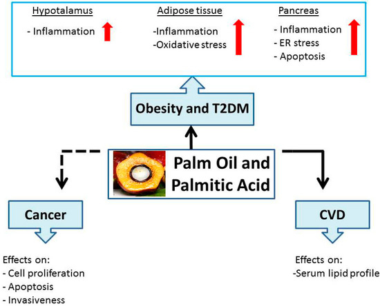
A schematic diagram representative of PO and PA effects on health, discussed in this review, is summarized in Figure 3.
Figure 3. Schematic representation of palm oil and palmitic acid effects on human health.

Acknowledgments

Grants from CEINGE Regione Campania (DGRC 1901/2009), IRCCS SDN, Ministry of Health, POR Campania FSE 2007-2013-CUP B25B09000050007, Project CREME, and Project PON02_00619_3461281.

Author Contributions

Pasqualina Buono developed the concept of the manuscript and performed the final revision; Concetta Montagnese contributed to the Introduction section; Annamaria Mancini, Aurora Daniele, and Ersilia Nigro contributed to the role of PO in obesity and T2DM; Stefania Orrù contributed to the role of PO in CVD; Esther Imperlini contributed to the role of PO in cancer.

Conflicts of Interest

The authors declare no conflict of interest.

References

  1. Aranceta, J.; Perez-Rodrigo, C. Recommended dietary reference intakes, nutritional goals and dietary guidelines for fat and fatty acids: A systematic review. Br. J. Nutr. 2012107, S8–S22. [Google Scholar] [PubMed]
  2. Assmann, G.; Buono, P.; Daniele, A.; Della Valle, E.; Farinaro, E.; Ferns, G.; Krogh, V.; Kromhout, D.; Masana, L.; Merino, J.; et al. Functional foods and cardiometabolic diseases. International Task Force for Prevention of Cardiometabolic Diseases. Nutr. Metab. Cardiovasc. Dis. 201424, 1272–1300. [Google Scholar] [CrossRef] [PubMed][Green Version]
  3. Berger, N.A. Obesity and cancer pathogenesis. Ann. N. Y. Acad. Sci. 20141311, 57–76. [Google Scholar] [CrossRef] [PubMed]
  4. Mba, O.I.; Dumont, M.J.; Ngadi, M. Palm Oil: Processing, characterization and utilization in the food industry. A review. Food Biosci. 201510, 26–41. [Google Scholar] [CrossRef]
  5. Sambanthamurthi, R.; Sundram, K.; Tan, Y.A. Chemistry and biochemistry of Palm Oil. Prog. Lip Res. 200039, 507–558. [Google Scholar] [CrossRef]
  6. Obibuzor, J.U.; Okogbenin, E.A.; Abigor, R.D. Oil recovery from palm fruits and palm kernl. In Palm Oil: Production, Processing, Characterization and Uses; Lai, O.-M., Tan, C.-P., Akoh, C.C., Eds.; AOCS Press: Urbana, IL, USA, 2012; pp. 299–328. [Google Scholar]
  7. Edem, D.O. Palm Oil: Biochemical, physiological, nutritional, hematological, and toxicological aspects: A review. Plant. Foods Hum. Nutr. 200257, 319–341. [Google Scholar] [CrossRef] [PubMed]
  8. Sundram, K.; Sambanthamurthi, R.; Tan, Y.A. Palm fruit chemistry and nutrition. Asia Pac. J. Clin. Nutr. 200312, 355–362. [Google Scholar] [PubMed]
  9. Souganidis, E.; Laillou, A.; Leyvraz, M.; Moench-Pfanner, R. A comparison of retinyl palmitate and red Palm Oil β-carotene as strategies to address Vitamin A deficiency. Nutrients 201315, 3257–3271. [Google Scholar]
  10. Ong, A.S.; Goh, S.H. Palm Oil: A healthful and cost-effective dietary component. Food Nutr. Bull. 200223, 11–22. [Google Scholar] [PubMed]
  11. Sen, C.K.; Khanna, S.; Roy, S. Tocotrienols in health and disease: The other half of the natural vitamin E family. Mol. Asp. Med. 200728, 692–728. [Google Scholar] [CrossRef] [PubMed]
  12. Čmolík, J.; Pokorný, J. Physical refining of edible oils. Eur. J. Lipid Sci. Technol. 2000102, 472–486. [Google Scholar] [CrossRef]
  13. Dunford, N.T. Advancements in oil and oilseed processing. In Food and Industrial Bioproducts and Bioprocessing; John Wiley and Sons Inc.: Ames, IA, USA, 2012; pp. 115–143. [Google Scholar]
  14. Gunstone, F.D. Vegetable Oils in Food Technology: Composition, Properties and Uses, 2nd ed.; Blackwell Publishing Ltd.: West Essex, UK, 2011; pp. 25–133. [Google Scholar]
  15. Henson, I.E. A brief history of the palm oil. In Palm Oil: Production, Processing, Characterization and Uses; Lai, O.-M., Tan, C.-P., Akoh, C.C., Eds.; AOCS Press: Urbana, IL, USA, 2012. [Google Scholar]
  16. Gee, P.T. Analytical characteristics of crude and refined Palm Oil and fractions. Eur. J. Lipid Sci. Technol. 2007109, 373–379. [Google Scholar] [CrossRef]
  17. Jensen, R.G. The lipids in human milk. Prog. Lipid Res. 199635, 53–92. [Google Scholar] [CrossRef]
  18. Odia, O.J.; Ofori, S.; Maduka, O. Palm Oil and the heart: A review. World J. Cardiol. 201526, 144–149. [Google Scholar] [CrossRef]
  19. Fattore, E.; Fanelli, R. Palm oil and palmitic acid: A review on cardiovascular effects and carcinogenicity. Int. J. Food Sci. Nutr. 201364, 648–659. [Google Scholar] [CrossRef]
  20. Fattore, E.; Bosetti, C.; Brighenti, F.; Agostoni, C.; Fattore, G. Palm oil and blood lipid-related markers of cardiovascular disease: A systematic review and meta-analysis of dietary intervention trials. Am. J. Clin. Nutr. 201499, 1331–1350. [Google Scholar] [PubMed]
  21. Bester, D.; Esterhuyse, A.; Truter, E.J.; van Rooyen, J. Cardiovascular effects of edible oils: A comparison between four popular edible oils. Nutr. Res. Rev. 201023, 334–348. [Google Scholar] [CrossRef] [PubMed]
  22. May, C.Y.; Nesaretnam, K. Research advancements in palm oil nutrition. Eur. J. Lipid Sci. Technol. 2014116, 1301–1315. [Google Scholar] [PubMed]
  23. Dixon, J.B. The effect of obesity on health outcomes. Mol. Cell. Endocrinol. 2010316, 104–108. [Google Scholar] [CrossRef] [PubMed]
  24. World Health Organization. Obesity and Overweight. Available online: http://www.who.int/mediacentre/factsheets/fs311/en/ (accessed on 19 January 2015).
  25. Kopelman, P.G. Obesity as a medical problem. Nature 2000404, 635–643. [Google Scholar] [PubMed]
  26. Exley, M.A.; Hand, L.; O’Shea, D.; Lynch, L. Interplay between the immune system and adipose tissue in obesity. J. Endocrinol. 2014223, R41–R48. [Google Scholar] [CrossRef] [PubMed]
  27. Ajuwon, K.M.; Spurlock, M.E. Palmitate activates the NF-κB transcription factor and induces IL-6 and TNF-α expression in 3T3-L1 adipocytes. J. Nutr. 2005135, 1841–1846. [Google Scholar] [PubMed]
  28. Bradley, R.L.; Fisher, F.F.; Maratos-Flier, E. Dietary fatty acids differentially regulate production of TNF-α and IL-10 by murine 3T3-L1 adipocytes. Obesity 200816, 938–944. [Google Scholar] [CrossRef] [PubMed]
  29. Wang, X.; Cheng, M.; Zhao, M.; Ge, A.; Guo, F.; Zhang, M.; Yang, Y.; Liu, L.; Yang, N. Differential effects of high-fat-diet rich in lard oil or soybean oil on osteopontin expression and inflammation of adipose tissue in diet-induced obese rats. Eur. J. Nutr. 201352, 1181–1189. [Google Scholar] [PubMed]
  30. Ting, J.P.; Willingham, S.B.; Bergstralh, D.T. NLRs at the intersection of cell death and immunity. Nat. Rev. Immunol. 20088, 372–379. [Google Scholar] [CrossRef] [PubMed]
  31. Franchi, L.; Eigenbrod, T.; Munoz-Planillo, R.; Nunez, G. The inflammasome: A caspase-1-activation platform that regulates immune responses and disease pathogenesis. Nat. Immunol. 200910, 241–247. [Google Scholar] [PubMed]
  32. Jager, J.; Gremeaux, T.; Cormont, M.; Le Marchand-Brustel, Y.; Tanti, J.F. Interleukin-1β-induced insulin resistance in adipocytes through down-regulation of insulin receptor substrate-1 expression. Endocrinology 2007148, 241–251. [Google Scholar] [CrossRef] [PubMed][Green Version]
  33. Wen, H.; Gris, D.; Lei, Y.; Jha, S.; Zhang, L.; Huang, M.T.; Brickey, W.J.; Ting, J.P. Fatty acid-induced NLRP3-ASC inflammasome activation interferes with insulin signaling. Nat. Immunol. 201112, 408–415. [Google Scholar] [CrossRef] [PubMed]
  34. Cani, P.D.; Amar, J.; Iglesias, M.A.; Poggi, M.; Knauf, C.; Bastelica, D.; Neyrinck, A.M.; Fava, F.; Tuohy, K.M.; Chabo, C.; et al. Metabolic endotoxemia initiates obesity and insulin resistance. Diabetes 200756, 1761–1772. [Google Scholar] [PubMed]
  35. Cani, P.D.; Bibiloni, R.; Knauf, C.; Waget, A.; Neyrinck, A.M.; Delzenne, N.M.; Burcelin, R. Changes in gut microbiota control metabolic endotoxemiainduced inflammation in high-fat diet-induced obesity and diabetes in mice. Diabetes 200857, 1470–1481. [Google Scholar] [PubMed]
  36. Stoll, L.L.; Denning, G.M.; Li, W.G.; Rice, J.B.; Harrelson, A.L.; Romig, S.A.; Gunnlaugsson, S.T.; Miller, F.J., Jr.; Weintraub, N.L. Regulation of endotoxin-induced proinflammatory activation in human coronary artery cells: Expression of functional membrane-bound CD14 by human coronary artery smooth muscle cells. J. Immunol. 2004173, 1336–1343. [Google Scholar] [CrossRef] [PubMed]
  37. Ghoshal, S.; Witta, J.; Zhong, J.; De Villiers, W.; Eckhardt, E. Chylomicrons promote intestinal absorption of lipopolysaccharides. J. Lipid Res. 200950, 90–97. [Google Scholar] [CrossRef] [PubMed]
  38. Laugerette, F.; Vors, C.; Geloen, A.; Chauvin, M.A.; Soulage, C.; Lambert-Porcheron, S.; Peretti, N.; Alligier, M.; Burcelin, R.; Laville, M.; et al. Emulsified lipids increase endotoxemia: Possible role in early postprandial low-grade inflammation. J. Nutr. Biochem. 201122, 53–59. [Google Scholar] [CrossRef] [PubMed]
  39. Laugerette, F.; Furet, J.P.; Debard, C.; Daira, P.; Loizon, E.; Géloën, A.; Soulage, C.O.; Simonet, C.; Lefils-Lacourtablaise, J.; Bernoud-Hubac, N.; et al. Oil composition of high-fat diet affects metabolic inflammation differently in connection with endotoxin receptors in mice. Am. J. Physiol. Endocrinol. Metab. 2012302, E374–E386. [Google Scholar] [CrossRef] [PubMed][Green Version]
  40. Ley, R.E.; Bäckhed, F.; Turnbaugh, P.; Lozupone, C.A.; Knight, R.D.; Gordon, J.I. Obesity alters gut microbial ecology. Proc. Natl. Acad. Sci. USA 2005102, 11070–11075. [Google Scholar] [CrossRef] [PubMed]
  41. Musso, G.; Gambino, R.; Cassader, M. Interactions between gut microbiota and host metabolism predisposing to obesity and diabetes. Annu. Rev. Med. 201162, 361–380. [Google Scholar] [PubMed]
  42. De Wit, N.; Derrien, M.; Bosch-Vermeulen, H.; Oosterink, E.; Keshtkar, S.; Duval, C.; de Vogel-van den Bosch, J.; Kleerebezem, M.; Müller, M.; van der Meer, R. Saturated fat stimulates obesity and hepatic steatosis and affects gut microbiota composition by an enhanced overflow of dietary fat to the distal intestine. Am. J. Physiol. Gastrointest. Liver Physiol. 2012303, G589–G599. [Google Scholar] [CrossRef] [PubMed]
  43. Musa, C.V.; Mancini, A.; Alfieri, A.; Labruna, G.; Valerio, G.; Franzese, A.; Pasanisi, F.; Licenziati, M.R.; Sacchetti, L.; Buono, P. Four novel UCP3 gene variants associated with childhood obesity: Effect on fatty acid oxidation and on prevention of triglyceride storage. Int. J. Obes. 201236, 207–217. [Google Scholar] [CrossRef] [PubMed]
  44. Cheng, L.; Yu, Y.; Szabo, A.; Wu, Y.; Wang, H.; Camer, D.; Huang, X.F. Palmitic acid induces central leptin resistance and impairs hepatic glucose and lipid metabolism in male mice. J. Nutr. Biochem. 201526, 541–548. [Google Scholar] [CrossRef] [PubMed]
  45. Thompson, N.M.; Norman, A.M.; Donkin, S.S.; Shankar, R.R.; Vickers, M.H.; Miles, J.L. Prenatal and postnatal pathways to obesity: Different underlying mechanisms, different metabolic outcomes. Endocrinology 2007148, 2345–2354. [Google Scholar] [CrossRef] [PubMed]
  46. Barker, D.J. The developmental origins of adult disease. J. Am. Coll. Nutr. 200423, 588S–595S. [Google Scholar] [CrossRef] [PubMed]
  47. Silva, A.P.; Guimaraes, D.E.; Mizurini, D.M.; Maia, I.C.; Ortiz-Costa, S.; Sardinha, F.L. Dietary fatty acids early in life affect lipid metabolism and adiposity in young rats. Lipids 200641, 535–541. [Google Scholar] [CrossRef] [PubMed]
  48. Magri, T.P.; Fernandes, F.S.; Souza, A.S.; Langhi, L.G.; Barboza, T.; Misan, V.; Mucci, D.B.; Santos, R.M.; Nunes, T.F.; Souza, S.A.; et al. Interesterified fat or palmoil as substitutes for partially hydrogenated fat in maternal diet can predispose obesity in adult male offspring. Clin. Nutr. 201524, 904–910. [Google Scholar] [CrossRef]
  49. De Rosa, A.; Monaco, M.L.; Capasso, M.; Forestieri, P.; Pilone, V.; Nardelli, C.; Buono, P.; Daniele, A. Adiponectin oligomers as potential indicators of adipose tissue improvement in obese subjects. Eur. J. Endocrinol. 2013169, 37–43. [Google Scholar]
  50. Jiao, P.; Ma, J.; Feng, B.; Zhang, H.; Diehl, J.A.; Chin, Y.E.; Yan, W.; Xu, H. FFA-induced adipocyte inflammation and insulin resistance: Involvement of ER stress and IKKβ pathways. Obesity 201119, 483–491. [Google Scholar] [CrossRef] [PubMed]
  51. Kharroubi, A.T.; Darwish, H.M. Diabetes mellitus: The epidemic of the century. World J. Diabetes 20156, 850–867. [Google Scholar] [CrossRef] [PubMed]
  52. Daniele, A.; Cammarata, R.; Pasanisi, F.; Finelli, C.; Salvatori, G.; Calcagno, G.; Bracale, R.; Labruna, G.; Nardelli, C.; Buono, P.; et al. Molecular analysis of the adiponectin gene in severely obese patients from southern Italy. Ann. Nutr. Metab. 200853, 155–161. [Google Scholar] [CrossRef] [PubMed]
  53. Gallagher, E.J.; LeRoith, D. Obesity and Diabetes: The Increased Risk of Cancer and Cancer-Related Mortality. Physiol. Rev. 201595, 727–748. [Google Scholar] [CrossRef] [PubMed]
  54. Zhang, K. Integration of ER stress, oxidative stress and the inflammatory response in health and disease. Int. J. Clin. Exp. Med. 20103, 33–40. [Google Scholar] [PubMed]
  55. Kochikuzhyil, B.M.; Devi, K.; Fattepur, S.R. Effect of saturated fatty acid-rich dietary vegetable oils on lipid profile, antioxidant enzymes and glucose tolerance in diabetic rats. Indian J. Pharmacol. 201042, 142–145. [Google Scholar] [PubMed]
  56. Storlien, L.H.; Higgins, J.A.; Thomas, T.C.; Brown, M.A.; Wang, H.Q.; Huang, X.F.; Else, P.L. Diet composition and insulin action in animal models. Br. J. Nutr. 200083, S85–S90. [Google Scholar] [CrossRef] [PubMed]
  57. Ariyama, H.; Kono, N.; Matsuda, S.T.; Inoue, H. Decrease in membrane phospholipid unsaturation induces unfolded protein response. J. Biol. Chem. 2010285, 22027–22035. [Google Scholar] [CrossRef] [PubMed]
  58. Van Amelsvoort, J.M.; van der Beek, A.; Stam, J.J. Effects of the type of dietary fatty acid on the insulin receptor function in rat epididymal fat cells. Ann. Nutr. Metab. 198630, 273–280. [Google Scholar] [CrossRef] [PubMed]
  59. Kritchevsky, D.; Sundram, K. Palm oil in human nutrition: Recent advances. Asia Pac. J. Clin. Nutr. 200211 (Suppl. 7), S393. [Google Scholar] [CrossRef]
  60. Rosqvist, F.; Iggman, D.; Kullberg, J.; Cedernaes, J.; Johansson, H.E.; Larsson, A.; Johansson, L.; Ahlström, H.; Arner, P.; Dahlman, I.; et al. Overfeeding polyunsaturated and saturated fat causes distinct effects on liver and visceral fat accumulation in humans. Diabetes 201463, 2356–2368. [Google Scholar] [CrossRef] [PubMed]
  61. Forouhi, N.G.; Koulman, A.; Sharp, S.J.; Imamura, F.; Kröger, J.; Schulze, M.B. Differences in the prospective association between individual plasma phospholipid saturated fatty acids and incident type 2 diabetes: The EPIC-InterAct case-cohort study. Lancet Diabetes Endocrinol. 20142, 810–818. [Google Scholar] [CrossRef]
  62. Yang, M.; Wei, D.; Mo, C.; Zhang, J.; Wang, X.; Han, X.; Wang, Z.; Xiao, H. Saturated fatty acid palmitate-induced insulin resistance is accompanied with myotube loss and the impaired expression of health benefit myokine genes in C2C12 myotubes. Lipids Health Dis. 201312, 104–113. [Google Scholar] [CrossRef] [PubMed]
  63. Simon-Szabó, L.; Kokas, M.; Mandl, J.; Kéri, G.; Csala, M. Metformin Attenuates Palmitate-Induced Endoplasmic Reticulum Stress, Serine Phosphorylation of IRS-1 and Apoptosis in Rat Insulinoma Cells. PLoS ONE 20149, e97868–e97875. [Google Scholar] [CrossRef] [PubMed][Green Version]
  64. Wei, Y.; Wang, D.; Topczewski, F.; Passagliotti, M.J. Saturated fatty acids induce endoplasmic reticulum stress and apoptosis indepently of ceramide in liver cells. Am. J. Physiol. Endocrinol. Metabol. 2006291, E275–E281. [Google Scholar] [CrossRef] [PubMed]
  65. Diakogiannaki, E.; Welters, H.J.; Morgan, N.G. Differential regulation of the endoplasmic reticulum stress response in pancreatic beta-cells exposed to long-chain saturated and monounsaturated fatty acids. J. Endocrinol. 2008197, 553–563. [Google Scholar] [CrossRef] [PubMed]
  66. Mordier, S.; Iynedjian, P.B. Activation of mammalian target of rapamycin complex 1 and insulin resistance induced by palmitate in hepatocytes. Biochem. Biophys. Res. Commun. 2007362, 206–211. [Google Scholar] [CrossRef] [PubMed]
  67. Ishii, M.; Maeda, A.; Tani, S.; Akagawa, M. Palmitate induces insulin resistance in human HepG2 hepatocytes by enhancing ubiquitination and proteasomal degradation of key insulin signaling molecules. Arch. Biochem. Biophys. 2015566, 26–35. [Google Scholar] [CrossRef] [PubMed]
  68. Kronenberg, F.; Kronenberg, M.F.; Kiechl, S.; Trenkwalder, E.; Santer, P.; Oberhollenzer, F.; Egger, G.; Utermann, G.; Willeit, J. Role of lipoprotein(a) and apolipoprotein(a) phenotype in atherogenesis: Prospective results from the Bruneck study. Circulation 1999100, 1154–1160. [Google Scholar] [CrossRef] [PubMed]
  69. Walldius, G.; Jungner, I.; Holme, I.; Aastveit, A.H.; Kolar, W.; Steiner, E. High apolipoprotein B, low apolipoprotein A-I, and improvement in the prediction of fatal myocardial infarction (AMORIS study): A prospective study. Lancet 2001358, 2026–2033. [Google Scholar] [CrossRef]
  70. Walldius, G.; Jungner, I. Apolipoprotein B and apolipoprotein A-I: Risk indicators of coronary heart disease and targets for lipid-modifying therapy. J. Intern. Med. 2004255, 188–205. [Google Scholar] [CrossRef] [PubMed]
  71. Keys, A.; Menotti, A.; Aravanis, C.; Blackburn, H.; Djordevic, B.S.; Buzina, R.; Dontas, A.S.; Fidanza, F.; Karvonen, M.J.; Kimura, N.; et al. The seven countries study: 2289 deaths in 15 years. Prev. Med. 198413, 141–154. [Google Scholar] [CrossRef]
  72. Keys, A.; Menotti, A.; Karvonen, M.J.; Aravanis, C.; Blackburn, H.; Buzina, R.; Djordjevic, B.S.; Dontas, A.S.; Fidanza, F.; Keys, M.H.; et al. The diet and 15-year death rate in the seven countries study. Am. J. Epidemiol. 1986124, 903–915. [Google Scholar] [PubMed]
  73. Kromhout, D.; Keys, A.; Aravanis, C.; Buzina, R.; Fidanza, F.; Giampaoli, S.; Jansen, A.; Menotti, A.; Nedeljkovic, S.; Pekkarinen, M. Food consumption patterns in the 1960s in seven countries. Am. J. Clin. Nutr. 198949, 889–894. [Google Scholar] [PubMed]
  74. Menotti, A.; Keys, A.; Aravanis, C.; Blackburn, H.; Dontas, A.; Fidanza, F.; Karvonen, M.J.; Kromhout, D.; Nedeljkovic, S.; Nissinen, A.; Toshima, H.; et al. Seven countries study. First 20-year mortality data in 12 cohorts of six countries. Ann. Med. 198921, 175–179. [Google Scholar] [CrossRef] [PubMed]
  75. Verschuren, W.M.; Jacobs, D.R.; Bloemberg, B.P.; Kromhout, D.; Menotti, A.; Aravanis, C.; Blackburn, H.; Buzina, R.; Dontas, A.S.; Fidanza, F.; et al. Serum total cholesterol and long-term coronary heart disease mortality in different cultures. Twenty-five-year follow-up of the seven countries study. J. Am. Med. Assoc. 1995274, 131–136. [Google Scholar] [CrossRef]
  76. Mensink, R.P.; Katan, M.B. Effect of dietary fatty acids on serum lipids and lipoproteins. A meta-analysis of 27 trials. Arterioscler. Thromb. 199212, 911–919. [Google Scholar] [CrossRef] [PubMed]
  77. Mensink, R.P.; Zock, P.L.; Kester, A.D.; Katan, M.B. Effects of dietary fatty acids and carbohydrates on the ratio of serum total to HDL cholesterol and on serum lipids and apolipoproteins: A meta-analysis of 60 controlled trials. Am. J. Clin. Nutr. 200377, 1146–1155. [Google Scholar] [PubMed]
  78. Chen, B.K.; Seligman, B.; Farquhar, J.W.; Goldhaber-Fiebert, J.D. Multi-Country analysis of palm oil consumption and cardiovascular disease mortality for countries at different stages of economic development: 1980–1997. Glob. Health 20117, 45–54. [Google Scholar] [CrossRef] [PubMed]
  79. Utarwuthipong, T.; Komindr, S.; Pakpeankitvatana, V.; Songchitsomboon, S.; Thongmuang, N. Small dense low-density lipoprotein concentration and oxidative susceptibility changes after consumption of soybean oil, rice bran oil, palm oil and mixed rice bran/palm oil in hypercholesterolaemic women. J. Int. Med. Res. 200937, 96–104. [Google Scholar] [CrossRef]
  80. Truswell, A.S. Comparing palmolein with different predominantly monounsaturated oils: Effect on plasma lipids. Int. J. Food Sci. Nutr. 200051, S73–S77. [Google Scholar] [CrossRef] [PubMed]
  81. Vega-López, S.; Ausman, L.M.; Jalbert, S.M.; Erkkilä, A.T.; Lichtenstein, A.H. Palm and partially hydrogenated soybean oils adversely alter lipoprotein profiles compared with soybean and canola oils in moderately hyperlipidemic subjects. Am. J. Clin. Nutr. 200684, 54–62. [Google Scholar] [PubMed]
  82. Clandinin, M.T.; Cook, S.L.; Konrad, S.D.; Goh, Y.K.; French, M.A. The effect of palmitic acid on lipoprotein cholesterol levels and endogenous cholesterol synthesis in hyperlipidemic subjects. Lipids 199934, S121–S124. [Google Scholar] [CrossRef]
  83. Clandinin, M.T.; Cook, S.L.; Konrad, S.D.; French, M.A. The effect of palmitic acid on lipoprotein cholesterol levels. Int. J. Food Sci. Nutr. 200051, S61–S71. [Google Scholar] [PubMed]
  84. Marzuki, A.; Arshad, F.; Razak, T.A.; Jaarin, K. Influence of dietary fat on plasma lipid profiles of Malaysian adolescents. Am. J. Clin. Nutr. 199153 (Suppl. 4), 1010S–1014S. [Google Scholar] [PubMed]
  85. Sundram, K.; Ismail, A.; Hayes, K.C.; Jeyamalar, R.; Pathmanathan, R. Trans (elaidic) fatty acids adversely affect the lipoprotein profile relative to specific saturated fatty acids in humans. J. Nutr. 1997127, 514S–520S. [Google Scholar] [PubMed]
  86. Sundram, K.; Hayes, K.C.; Siru, O.H. Dietary palmitic acid results in lower serum cholesterol than does a lauric-myristic acid combination in normolipemic humans. Am. J. Clin. Nutr. 199459, 841–846. [Google Scholar] [PubMed]
  87. Favé, G.; Coste, T.C.; Armand, M. Physicochemical properties of lipids: New strategies to manage fatty acid bioavailability. Cell. Mol. Biol. 200450, 815–831. [Google Scholar] [PubMed]
  88. Karupaiah, T.; Sundram, K. Effects of stereospecific positioning of fatty acids in triacylglycerol structures in native and randomized fats: A review of their nutritional implications. Nutr. Metab. 20074, 16–32. [Google Scholar] [CrossRef] [PubMed]
  89. Kromhout, D.; Menotti, A.; Bloemberg, B.; Aravanis, A.; Blackburn, H.; Buzina, R.; Dontas, A.S.; Fidanza, F.; Giampaoli, S.; Jansen, A. Dietary saturated and trans fatty acids and cholesterol and 25-year mortality from coronary heart disease: The Seven Countries Study. Prev. Med. 199524, 308–315. [Google Scholar] [CrossRef] [PubMed]
  90. Clarke, R.; Frost, C.; Collins, R.; Appleby, P.; Peto, R. Dietary lipids and blood cholesterol: Quantitative meta-analysis of metabolic ward studies. BMJ 1997314, 112–117. [Google Scholar] [CrossRef] [PubMed]
  91. Renaud, S.C.; Ruf, J.C.; Petithory, D. The positional distribution of fatty acids in palm oil and lard influences their biologic effects in rats. J. Nutr. 1995125, 229–237. [Google Scholar]
  92. Kritchevsky, D.; Tepper, S.A.; Wright, S.; Kuksis, A.; Hughes, T.A. Cholesterol vehicle in experimental atherosclerosis. Cottonseed oil and randomized cottonseed oil. Nutr. Res. 199818, 259–264. [Google Scholar] [CrossRef]
  93. Kritchevsky, D.; Tepper, S.A.; Kuksis, A.; Eghtedary, K.; Klurfeld, D.M. Cholesterol vehicle in experimental atherosclerosis. Native and randomized lard and tallow. J. Nutr. Biochem. 19989, 582–585. [Google Scholar] [CrossRef]
  94. Kritchevsky, D.; Tepper, S.A.; Chen, S.C.; Meijer, G.W.; Krauss, R.M. Cholesterol vehicle in experimental atherosclerosis. 23. Effects of specific synthetic triglycerides. Lipids 200035, 621–625. [Google Scholar] [CrossRef] [PubMed]
  95. Kritchevsky, D.; Tepper, S.A.; Czarnecki, S.K.; Sundram, K. Red palm oil in experimental atherosclerosis. Asia Pacific J. Clin. Nutr. 200211, S433–S437. [Google Scholar] [CrossRef]
  96. López-López, A.; Castellote-Bargalló, A.I.; Campoy-Folgoso, C.; Rivero-Urgël, M.; Tormo-Carnicé, R.; Infante-Pina, D.; López-Sabater, M.C. The influence of dietary palmitic acid triacylglyceride position on the fatty acid, calcium and magnesium contents of at term newborn faeces. Early Hum. Dev. 200165, S83–S94. [Google Scholar] [CrossRef]
  97. Sczaniecka, A.K.; Brasky, T.M.; Lampe, J.W.; Patterson, R.E.; White, E. Dietary intake of specific fatty acids and breast cancer risk among postmenopausal women in the VITAL cohort. Nutr. Cancer 201264, 1131–1142. [Google Scholar] [CrossRef] [PubMed]
  98. Hodge, A.M.; Williamson, E.J.; Bassett, J.K.; MacInnis, R.J.; Giles, G.G.; English, D.R. Dietary and biomarker estimates of fatty acids and risk of colorectal cancer. Int. J. Cancer 2015. [Google Scholar] [CrossRef] [PubMed]
  99. Jackson, M.D.; Walker, S.P.; Simpson-Smith, C.M.; Lindsay, C.M.; Smith, G.; McFarlane-Anderson, N.; Bennett, F.I.; Coard, K.C.; Aiken, W.D.; Tulloch, T.; et al. Associations of whole-blood fatty acids and dietary intakes with prostate cancer in Jamaica. Cancer Causes Control 201223, 23–33. [Google Scholar] [CrossRef] [PubMed]
  100. Saadatian-Elahi, M.; Norat, T.; Goudable, J.; Riboli, E. Biomarkers of dietary fatty acid intake and the risk of breast cancer: A meta-analysis. Int. J. Cancer 2004111, 584–591. [Google Scholar] [CrossRef] [PubMed]
  101. Shen, X.J.; Zhou, J.D.; Dong, J.Y.; Ding, W.Q.; Wu, J.C. Dietary intake of n-3 fatty acids and colorectal cancer risk: A meta-analysis of data from 489,000 individuals. Br. J. Nutr. 2012108, 1550–1556. [Google Scholar] [CrossRef] [PubMed]
  102. Brouwer, I.A.; Katan, M.B.; Zock, P.L. Dietary α-linolenic acidis associated with reduced risk of fatal coronary heart disease, but increased prostate cancer risk: A meta-analysis. J. Nutr. 2004134, 919–922. [Google Scholar] [PubMed]
  103. Wolk, A.; Bergstrom, R.; Hunter, D.; Willett, W.; Ljung, H.; Holmberg, L.; Bergkvist, L.; Bruce, A.; Adami, H.O. A prospective study of association of monounsaturated fat and other types of fat with risk of breast cancer. Arch. Intern. Med. 1998158, 41–45. [Google Scholar] [CrossRef] [PubMed]
  104. Lof, M.; Sandin, S.; Lagiou, P.; Hilakivi-Clarke, L.; Trichopoulos, D.; Adami, H.O.; Weiderpass, E. Dietary fat and breast cancer risk in the Swedish women’s lifestyle and health cohort. Br. J. Cancer 200797, 1570–1576. [Google Scholar] [CrossRef] [PubMed]
  105. Sieri, S.; Krogh, V.; Ferrari, P.; Berrino, F.; Pala, V.; Thiébaut, A.C.; Tjønneland, A.; Olsen, A.; Overvad, K.; Jakobsen, M.U. Dietary fat and breast cancer risk in the European Prospective Investigation into Cancer and Nutrition. Am. J. Clin. Nutr. 200888, 1304–1312. [Google Scholar] [PubMed]
  106. Thiebaut, A.C.; Kipnis, V.; Chang, S.C.; Subar, A.F.; Thompson, F.E.; Rosenberg, P.S.; Hollenbeck, A.R.; Leitzmann, M.; Schatzkin, A. Dietary fat and postmenopausal invasive breast cancer in the national institutes of health-AARP diet and health study cohort. J. Natl. Cancer Inst. 200799, 451–462. [Google Scholar] [CrossRef] [PubMed]
  107. Theodoratou, E.; McNeill, G.; Cetnarskyj, R.; Farrington, S.M.; Tenesa, A.; Barnetson, R.; Porteous, M.; Dunlop, M.; Campbell, H. Dietary fatty acids and colorectal cancer: A case-control study. Am. J. Epidemiol. 2007166, 181–195. [Google Scholar] [CrossRef] [PubMed]
  108. Crowe, F.L.; Allen, N.E.; Appleby, P.N.; Overvad, K.; Aardestrup, I.V.; Johnsen, N.F.; Tjønneland, A.; Linseisen, J.; Kaaks, R.; Boeing, H.; et al. Fatty acid composition of plasma phospholipids and risk of prostate cancer in a case-control analysis nested within the European Prospective Investigation into Cancer and Nutrition. Am. J. Clin. Nutr. 200888, 1353–1363. [Google Scholar] [PubMed]
  109. Kurahashi, N.; Inoue, M.; Iwasaki, M.; Sasazuki, S.; Tsugane, A.S. Japan Public Health Center-Based Prospective Study Group. Dairy product, saturated fatty acid, and calcium intake and prostate cancer in a prospective cohort of Japanese men. Cancer Epidemiol. Biomark. Prev. 200817, 930–937. [Google Scholar] [CrossRef] [PubMed]
  110. Hardy, S.; Langelier, Y.; Prentki, M. Oleate activates phosphatidylinositol 3-kinase and promotes proliferation and reduces apoptosis of MDA-MB-231 breast cancer cells, whereas palmitate has opposite effects. Cancer Res. 200060, 6353–6358. [Google Scholar] [PubMed]
  111. Rossini, A.; Zanobbio, L.; Sfondrini, L.; Cavalleri, A.; Secreto, G.; Morelli, D.; Palazzo, M.; Sommariva, M.; Tagliabue, E.; Rumio, C.; et al. Influence of fatty acid-free diet on mammary tumor development and growth rate in HER-2/Neu transgenic mice. J. Cell. Physiol. 2013228, 242–249. [Google Scholar] [CrossRef] [PubMed]
  112. Bartsch, H.; Nair, J.; Owen, R.W. Dietary polyunsaturated fatty acids and cancers of the breast and colorectum: Emerging evidence for their role as risk modifiers. Carcinogenesis 199920, 2209–2218. [Google Scholar] [CrossRef] [PubMed]
  113. Kolonel, L.N.; Nomura, A.M.; Cooney, R.V. Dietary fat and prostate cancer: Current status. J. Natl. Cancer Inst. 199991, 414–428. [Google Scholar] [CrossRef] [PubMed]
  114. Escrich, E.; Solanas, M.; Moral, R.; Costa, I.; Grau, L. Are the olive oil and other dietary lipids related to cancer? Experimental evidence. Clin. Transl. Oncol. 20068, 868–883. [Google Scholar] [CrossRef] [PubMed]
  115. Kuriki, K.; Wakai, K.; Hirose, K.; Matsuo, K.; Ito, H.; Suzuki, T.; Saito, T.; Kanemitsu, Y.; Hirai, T.; Kato, T.; et al. Risk of colorectal cancer is linked to erythrocyte compositions of fatty acids as biomarkers for dietary intakes of fish, fat, and fatty acids. Cancer Epidemiol. Biomark. Prev. 200615, 1791–1798. [Google Scholar] [CrossRef] [PubMed]
  116. Shannon, J.; King, I.B.; Moshofsky, R.; Lampe, J.W.; Gao, D.L.; Ray, R.M.; Thomas, D.B. Erythrocyte fatty acids and breast cancer risk: A case-control study in Shanghai, China. Am. J. Clin. Nutr. 200785, 1090–1097. [Google Scholar] [PubMed]
  117. Escrich, E.; Solanas, M.; Moral, R.; Escrich, R. Modulatory effects and molecular mechanisms of olive oil and other dietary lipids in breast cancer. Curr. Pharm. Des. 201117, 813–830. [Google Scholar] [CrossRef] [PubMed]
  118. Calder, P.C. Dietary fatty acids and the immune system. Lipids 199934, S137–S140. [Google Scholar] [CrossRef] [PubMed]
  119. Ge, G.; Wu, J.; Lin, Q. Effect of membrane fluidity on tyrosine ki-nase activity of reconstituted epidermal growth factor receptor. Biochem. Biophys. Res. Commun. 2001282, 511–514. [Google Scholar] [CrossRef] [PubMed]
  120. Soto-Guzman, A.; Navarro-Tito, N.; Castro-Sanchez, L.; Martinez-Orozco, R.; Salazar, E.P. Oleic acid promotes MMP-9 secretion and invasion in breast cancer cells. Clin. Exp. Metastasis 201027, 505–515. [Google Scholar] [CrossRef] [PubMed]
  121. Matsuoka, T.; Adair, J.E.; Lih, F.B.; Hsi, L.C.; Rubino, M.; Eling, T.E.; Tomer, K.B.; Yashiro, M.; Hirakawa, K.; Olden, K.; et al. Elevated dietary linoleic acid increases gastric carcinoma cell invasion and metastasis in mice. Br. J. Cancer 2010103, 1182–1191. [Google Scholar] [CrossRef] [PubMed]
  122. Kuhajda, F.P.; Pizer, E.S.; Li, J.N.; Mani, N.S.; Frehywot, G.L.; Townsend, C.A. Synthesis and antitumor activity of an inhibitor of fatty acid synthase. Proc. Natl. Acad. Sci. USA 200097, 3450–3454. [Google Scholar] [CrossRef] [PubMed]
  123. Nigro, E.; Imperlini, E.; Scudiero, O.; Monaco, M.L.; Polito, R.; Mazzarella, G.; Orrù, S.; Bianco, A.; Daniele, A. Differentially expressed and activated proteins associated with non small cell lung cancer tissues. Respir. Res. 201516, 74. [Google Scholar] [CrossRef] [PubMed]
  124. Pandey, P.R.; Liu, W.; Xing, F.; Fukuda, K.; Watabe, K. Anti-cancer drugs targeting fatty acid synthase (FAS). Recent Pat. Anticancer Drug Discov. 20127, 185–197. [Google Scholar] [CrossRef] [PubMed]
  125. Liu, K.; Czaja, M.J. Regulation of lipid stores and metabolism by lipophagy. Cell. Death Differ. 201320, 3–11. [Google Scholar] [CrossRef] [PubMed]
  126. Knævelsrud, H.; Simonsen, A. Lipids in autophagy: Constituents, signaling molecules and cargo with relevance to disease. Biochim. Biophys. Acta 20121821, 1133–1145. [Google Scholar] [CrossRef] [PubMed]
  127. Singh, R.; Cuervo, A.M. Lipophagy: Connecting autophagy and lipid metabolism. Int. J. Cell Biol. 20122012. [Google Scholar] [CrossRef] [PubMed]
  128. Borradaile, N.M.; Han, X.; Harp, J.D.; Gale, S.E.; Ory, D.S.; Schaffer, J.E. Disruption of endoplasmic reticulum structure and integrity in lipotoxic cell death. J. Lipid Res. 200647, 2726–2737. [Google Scholar] [CrossRef] [PubMed]
  129. Cao, J.; Dai, D.L.; Yao, L.; Yu, H.H.; Ning, B.; Zhang, Q.; Chen, J.; Cheng, W.H.; Shen, W.; Yang, Z.X. Saturated fatty acid induction of endoplasmic reticulum stress and apoptosis in human liver cells via the PERK/ATF4/CHOP signaling pathway. Mol. Cell. Biochem. 2012364, 115–129. [Google Scholar] [CrossRef] [PubMed]
  130. Gu, X.; Li, K.; Laybutt, D.R.; He, M.L.; Zhao, H.L.; Chan, J.C.; Xu, G. Bip overexpression, but not CHOP inhibition, attenuates fatty-acid-induced endoplasmic reticulum stress and apoptosis in HepG2 liver cells. Life Sci. 201087, 724–732. [Google Scholar] [CrossRef] [PubMed]
  131. Park, E.J.; Lee, A.Y.; Park, S.; Kim, J.H.; Cho, M.H. Multiple pathways are involved in palmitic acid-induced toxicity. Food Chem. Toxicol. 201467, 26–34. [Google Scholar] [CrossRef] [PubMed]
  132. Unger, R.H.; Zhou, Y.T. Lipotoxicity of beta-cells in obesity and in other causes of fatty acid spillover. Diabetes 200150 (Suppl. 1), S118–S121. [Google Scholar] [CrossRef] [PubMed]
  133. Lambertucci, R.H.; Leandro, C.G.; Vinolo, M.A.; Nachbar, R.T.; Dos Reis Silveira, L.; Hirabara, S.M.; Curi, R.; Pithon-Curi, T.C. The effects of palmitic acid on nitric oxide production by rat skeletal muscle: Mechanism via superoxide and iNOS activation. Cell. Physiol. Biochem. 201230, 1169–1180. [Google Scholar] [CrossRef] [PubMed][Green Version]
  134. Hirabara, S.M.; Curi, R.; Maechler, P. Saturated fatty acid-induced insulin resistance is associated with mitochondrial dysfunction in skeletal muscle cells. J. Cell. Physiol. 2010222, 187–194. [Google Scholar] [CrossRef] [PubMed]
  135. Yuzefovych, L.; Wilson, G.; Rachek, L. Different effects of oleate vs. palmitate on mitochondrial function, apoptosis, and insulin signaling in L6 skeletal muscle cells: Role of oxidative stress. Am. J. Physiol. Endocrinol. Metab. 2010299, E1096–E1105. [Google Scholar] [CrossRef] [PubMed]
  136. Fromenty, B.; Robin, M.A.; Igoudjil, A.; Mansouri, A.; Pessayre, D. The ins and outs of mitochondrial dysfunction in NASH. Diabetes Metab. 200430, 121–38. [Google Scholar] [CrossRef]
  137. Nakamura, S.; Takamura, T.; Matsuzawa-Nagata, N.; Takayama, H.; Misu, H.; Noda, H.; Nabemoto, S.; Kurita, S.; Ota, T.; Ando, H.; Miyamoto, K.; et al. Palmitate induces insulin resistance in H4IIEC3 hepatocytes through reactive oxygen species produced by mitochondria. J. Biol. Chem. 2009284, 14809–14818. [Google Scholar] [CrossRef] [PubMed]
  138. Brookheart, R.T.; Michel, C.I.; Listenberger, L.L.; Ory, D.S.; Schaffer, J.E. The non-coding RNA gadd7 is a regulator of lipid-induced oxidative and endoplasmic reticulum stress. J. Biol. Chem. 2009284, 7446–7454. [Google Scholar] [CrossRef] [PubMed]
  139. Lee, Y.; Naseem, R.H.; Park, B.H.; Garry, D.J.; Richardson, J.A.; Schaffer, J.E.; Unger, R.H. Alpha-lipoic acid prevents lipotoxic cardiomyopathy in acyl CoA-synthase transgenic mice. Biochem. Biophys. Res. Commun. 2006344, 446–452. [Google Scholar] [CrossRef] [PubMed]
  140. Ye, G.; Metreveli, N.S.; Donthi, R.V.; Xia, S.; Xu, M.; Carlson, E.C.; Epstein, P.N. Catalase protects cardiomyocyte function in models of type 1 and type 2 diabetes. Diabetes 200453, 1336–1343. [Google Scholar] [CrossRef]
  141. Listenberger, L.L.; Ory, D.S.; Schaffer, J.E. Palmitate-induced apoptosis can occur through a ceramide-independent pathway. J. Biol. Chem. 2001276, 14890–14895. [Google Scholar] [CrossRef] [PubMed]
  142. García-Ruiz, I.; Fernández-Moreira, D.; Solís-Muñoz, P.; Rodríguez-Juan, C.; Díaz-Sanjuán, T.; Muñoz-Yagüe, T.; Solís-Herruzo, J.A. Mitochondrial complex I subunits are decreased in murine nonalcoholic fatty liver disease: Implication of peroxynitrite. J. Proteome Res. 20109, 2450–2459. [Google Scholar] [CrossRef] [PubMed]
  143. García-Ruiz, I.; Solís-Muñoz, P.; Fernández-Moreira, D.; Muñoz-Yagüe, T.; Solís-Herruzo, J.A. In vitro treatment of HepG2 cells with saturated fatty acids reproduces mitochondrial dysfunction found in nonalcoholic steatohepatitis. Dis. Model Mech. 20158, 183–191. [Google Scholar] [CrossRef] [PubMed]
  144. Khan, M.J.; Rizwan Alam, M.; Waldeck-Weiermair, M.; Karsten, F.; Groschner, L.; Riederer, M.; Hallström, S.; Rockenfeller, P.; Konya, V.; Heinemann, A.; et al. Inhibition of autophagy rescues palmitic acid-induced necroptosis of endothelial cells. J. Biol. Chem. 2012287, 21110–21120. [Google Scholar] [CrossRef] [PubMed]
$$$ If you are interested in a writer or editor position, check out here.We are hiring. $$$

20

No Responses

Write a response

five − two =