中文版谷歌中文翻譯(90% 準確率) | English translation
Buy/Sell Your Domains Here。在這裡購買/出售您的域名
Contact Dr. Lu for information about cancer treatments。聯繫盧博士,獲取有關癌症治療資訊。
Summary: The authors first isolated two active compounds from carrot and then incorporated them in certain dosage into feed intended for 20 study rats. 20 other rats were fed on a diet without the active compounds. All rats were exposed to a potent carcinogen and then entered the feeding trial. After 18 weeks, all were euthaized. Micro-tumors were found in 15 control rats (75%) and 8 study rats (40%) while big tumors in 6 control rats (40%) and 1 study rat (5%).
A few things to consider: 1) Carrot compounds did not render 100% prevention in this study. 2) Carrot compounds obviously inhibit the growth of tumor cells but the actual modes of anticancer mechanisms remain unknown.
總結:作者首先從胡蘿蔔中分離出兩種活性化合物,然後將它們按一定劑量加入用於 20 只研究大鼠的飼料中。 其他 20 隻大鼠以不含活性化合物的飲食餵養。 所有大鼠都暴露於強致癌物,然後進入餵養試驗。 18週後,所有人都被安樂死。 在 15 只對照大鼠 (75%) 和 8 只研究大鼠 (40%) 中發現了微腫瘤,而在 6 只對照大鼠 (40%) 和 1 只研究大鼠 (5%) 中發現了大腫瘤。
需要考慮的幾件事:1) 在這項研究中,胡蘿蔔化合物沒有提供 100% 的預防。 2)胡蘿蔔化合物明顯抑制腫瘤細胞的生長,但其抗癌機制的實際模式尚不清楚。
Read another report from the same university which is an observational study indicating that eating carrots can significantly reduce risk of colon cancer.
DOI: 10.1039/C7FO00110J (Paper) Food Funct., 2017, 8, 964-974
Open Access Article
This Open Access Article is licensed under a
Creative Commons Attribution 3.0 Unported Licence
DOI: 10.1039/C7FO00110J (Paper) Food Funct., 2017, 8, 964-974
Morten Kobaek-Larsen *ab, Rime B. El-Houri c, Lars P. Christensen c, Issam Al-Najami ab, Xavier Fretté c and Gunnar Baatrup ab
aDepartment of Clinical Research, University of Southern Denmark, Winsløwparken 19, DK-5000 Odense C, Denmark
bDepartment of Surgery A, Odense University Hospital, Valdemarsgade 53, DK-5700 Svendborg, Denmark. E-mail: [email protected]; Fax: +45 65919872; Tel: +45 30579310
cDepartment of Chemical Engineering, Biotechnology and Environmental Technology, University of Southern Denmark, Campusvej 55, DK-5230 Odense M, Denmark Received 20th January 2017 , Accepted 8th February 2017
First published on 9th February 2017
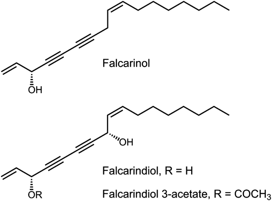
Falcarinol (FaOH) and falcarindiol (FaDOH) are found in many food plants of the Apiaceae family. Carrots are a major dietary source of these polyacetylenes. Feeding azoxymethane (AOM)-induced rats with carrots and purified FaOH have previously been shown to inhibit neoplastic transformations in the colon. FaOH and FaDOH have also shown to have a synergistic effect in vitro, resulting in a significant increased cytotoxic activity. Based on these findings the antineoplastic effect of FaOH and FaDOH (purity > 99%) was investigated in the AOM-induced rat model. Twenty rats received rat diet containing 7 μg FaOH per g feed and 7 μg FaDOH per g feed and 20 rats were controls receiving only rat diet. Then carcinogenesis was induced in all 40 rats with the carcinogen AOM. All animals received the designated diet for 2 weeks before AOM induction and continued on the designated diet throughout the experiment. Rats were euthanized 18 weeks after the first AOM injection and macroscopic polyp/cancers were measured, harvested and stained for histology. The difference in sizes of aberrant crypt foci (ACF) were analysed in a Wilcoxon rank sum test, in which the median number of small ACF was 218 in controls and 145 in polyacetylene treated rats (P < 0.001). Fifteen control rats and 8 treated rats had macroscopic tumors (P = 0.027). The number of tumors larger than 3 mm were 6 and 1 in control and treated rats, respectively (P = 0.032). In conclusion dietary supplements with FaOH and FaDOH reduced the number of neoplastic lesions as well as the growth rate of the polyps suggesting a preventive effect of FaOH and FaDOH on the development of colorectal cancer.
Carrots are the major source of α- and -β-carotene in the diet in Northern Europe and North America,1 and epidemiologic evidence has indicated that diets rich in carotenoid-rich fruits and vegetables, and high serum levels of β-carotene, are associated with a reduced risk of cancers.2 The link between a high intake of carrots and other vegetables rich in β-carotene, and a decreased risk of cancer disease has been interpreted as a likely preventive effect of this carotenoid.2,3 However, subsequent intervention studies have ruled out this explanation, since supplementation with β-carotene in well-nourished populations does not reduce the cancer incidence.2,4–10 In fact β-carotene supplementation seems to increase rather than decrease lung cancer incidence in smokers.2,4–7 β-Carotene could, however, be a biomarker for other nutraceuticals with a potential anticancer effect.
Carrots are also a major dietary source of structurally related C17-polyacetylenes of which falcarinol (FaOH), falcarindiol (FaDOH) and falcarindiol 3-acetate (FaDOH3Ac) [Fig. 1] consistently occur in highest concentrations.11–15 Among these FaDOH is usually the most abundant followed by FaOH.12,15–18 Falcarinol type polyacetylenes have shown many interesting bioactivities including antiinflammatory,13,16,17 antiplatelet-aggregatory,13,17,19–21 cytotoxic,11,13,17,22–31 and antitumor activity.24,32
The antiproliferative effect of FaOH, FaDOH and FaDOH3Ac on cancer
cells is probably related to their ability to arrest the cell cycle
progression at G2/M or other phases of the cell cycles leading to the
induction of apoptosis.26,28,30,31,33
The exact mechanisms of action of falcarinol type polyacetylenes in
cancer cells are, however, not known, but may be related to their
alkylating properties as demonstrated recently by Heydenreuter et al.,34 which showed that FaOH is able to inhibit enzymes via covalent alkylation. This is also in accordance with FaOH being a strong contact allergen35
and thus may be a potent immune-stimulator. The presence of a secondary
alcohol group at C-3 seems to be important for the alkylating
properties of FaOH due to the possibility to generate a reactive
resonance stabilized carbocation by the loss of water, which then
readily can react with biomolecules involved in, e.g.,
inflammation and cell proliferation such as the nuclear factor
kappa-light-chain-enhancer of activated B cells (NF-κB) signalling
pathway, cyclooxygenase (COX)-1 and -2, lipoxygenases (LOXs) and
cytokines,13,17,20,29,31 and could explain its possible effect on cell cycle arrest. This is in accordance with a study by Purup et al.,29
which showed that FaOH significantly inhibited cell proliferation in
human epithelial colorectal adenocarcinoma cells (Caco-2) at 2.5 μg mL−1, whereas its oxidized form, falcarinone [(Z)-1,9-heptadecadiene-4,6-diyn-3-one] only inhibited proliferation in Caco-2 cells at 20 μg mL−1. In addition FaOH appears to be more cytotoxic than FaDOH13,17,24,27,29,31 as for example demonstrated by Zidorn et al.27
who tested FaOH and FaDOH against five different cancer cell lines,
including the colorectal carcinoma cell lines HRT-18 and HT-29. The
bioactivity of FaDOH3Ac has only been studied in few investigations. In
leukemia cell lines it has been demonstrated that FaDOH3Ac was more
cytotoxic than FaOH followed by FaDOH,31 whereas in Caco-2 cells FaOH seem to be the most potent of these polyacetylenes.29 On the other hand FaDOH is a more potent antiinflammatory agent compared to FaOH and FaDOH3Ac;16
thus the bioactivity of falcarinol type polyacetylenes depends on many
factors, although they may have similar mechanisms of action.
The antiproliferative effect of falcarinol type polyacetylenes clearly depends on the cell lines.22,29,31 Furthermore, they may work synergistically as demonstrated on the proliferation of Caco-2 cells in vitro for FaOH and FaDOH.29 By keeping FaOH constant at 1 μg mL−1 and incubating Caco-2 cells and normal human intestinal epithelial cells (FHs 74 Int.) with FaOH and FaDOH in different ratios, a clear synergistic response on the inhibitory effect of cell proliferation was observed by adding FaDOH in 1, 5 and 10 times the concentration of FaOH.29 A similar synergistic response on the inhibitory effect of cell proliferation was also observed for carrot extracts with different proportions of FaOH, FaDOH and FaDOH3Ac.29 The results indicated that FaDOH in low doses may have very potent inhibitory effects on the proliferation of Caco-2 cells as well as other cell types when found in combination with low doses of FaOH. A similar synergistic effect may be expected for FaDOH3Ac and other falcarinol type polyacetylenes; thus synergistic interactions could be important for their anticancer activity.
Feeding azoxymethane (AOM)-induced rats with carrots and purified FaOH from carrots have been shown to inhibit neoplastic transformations in the colon.32 The potential anticancer activity of carrots and FaOH was demonstrated by feeding with carrot or purified FaOH in amounts corresponding to a daily human intake of 400–600 g fresh weight (FW) of carrot.32 Despite of the low concentrations of FaOH in the rat feed (3.5 μg g−1 feed) and only 8 rats in each group, dietary treatments with carrots and FaOH, respectively, showed a significant effect (P = 0.028) to reduce the number of neoplastic lesions with increasing size of lesion from no difference of the smallest lesions to a one-third reduction for the fully developed tumors.32
Colorectal cancer is the third most common type of cancer and accounts for approximately 10% of all cancer related deaths.36 Environmental factors, particularly diet, play roles in the development of this disease.37 FaOH and FaDOH are not only the most abundant polyacetylenes in carrots but also in many other vegetables of the Apiaceae family, including among others celery, celeriac, and parsnip, in contrast to FaDOH3Ac, which has so far only been detected in carrots;13,17 thus FaOH and FaDOH are considered to be the most important dietary polyacetylenes. Consequently, the aim of this study was to investigate the inhibitory effect of purified FaOH and FaDOH in a combined treatment in AOM-induced rats, in a ratio where these polyacetylenes have shown synergistic inhibitory effect on the proliferation of Caco-2 cells, and thus to verify earlier observations demonstrating a potential preventive effect of dietary polyacetylenes of the falcarinol type on the development of colorectal cancer.
CHROMASOLV® high-performance liquid chromatography (HPLC) grade of acetonitrile (ACN), ethyl acetate (EtOAc), ethanol (EtOH, 96%), formic acid, methanol, n-hexane and trifluoroacetic acid (TFA) were purchased from Sigma-Aldrich (Steinheim, Germany). Anhydrous sodium sulphate was obtained from Sigma-Aldrich and silica gel 60 aluminium sheets for thin-layer chromatography (TLC) and silica gel 60 (particle size 0.063–0.2 mm) for column chromatography were purchased from Merck (Darmstadt, Germany). Water was ultrapure generated with a Milli-Q system consisting of an Ultra Clear Basic UV system (Holm & Halby, Brøndby, Denmark). Azoxymethane (AOM) was purchased from Sigma-Aldrich (A5486) and diluted with sterile 0.9% NaCl to a concentration of 5 mg mL−1 at the Central Pharmacy of the Odense University Hospital (Odense, Denmark).
Carrots (Daucus carota ssp. sativus cv. Miami) were used for extraction of FaOH and FaDOH. The carrots were obtained from the nursery Vostrup Øko (Vostrup, Tarm, Denmark) and stored in a cold storage room with automatic regulation of the temperature at 5 ± 0.5 °C until use. FaOH and FaDOH were isolated from carrots according to the procedure described by El-Houri et al.38 with a few modifications. Sixty kilograms of shredded carrot roots were extracted twice with 25 L of EtOAc for 24 h at 22 °C in the dark with periodical shaking. The combined extracts were filtered and dried over anhydrous sodium sulphate, and concentrated in vacuo (30 °C) under dim light yielding a combined extract of approximately 84 g. Analysis of the extract by analytical HPLC14,38 revealed the presence of FaOH (6.7 mg g−1 extract) and FaDOH (21.1 mg g−1 extract) as the main constituents followed by FaDOH3Ac (1.7 mg g−1 extract). The extract was divided into 3 portions of approximately 28 g, and each portion of the extract was chromatographed by flash column chromatography under dim light conditions (400 g, silica gel 60), eluting with 100% n-hexane (600 mL), n-hexane–EtOAc (v/v) (9
![[thin space (1/6-em)]](https://www.rsc.org/images/entities/char_2009.gif)
:
1, 4
:
1, 7
:
3, 3
:
2, 1
:
1, 2
:
3, 3
:
7, 1
:
4, 1
:
9, 600 mL for each step), 100% ACN (1000 mL), and finally with methanol (1000 mL). Fractions were analysed by TLC and analytical HPLC14,38 and fractions containing crude FaOH and FaDOH were combined and further purified by preparative reversed phase (RP)-HPLC on a Dionex UltiMate 3000 Binary Semi-preparative LC System from Thermo Fisher Scientific (Hvidovre, Denmark), equipped with a photodiode array detector (PDA), and a Foxy Jr. fraction collector unit from Teledyne ISCO Inc. (NE, USA). Separations were performed on a Develosil ODS-HG-5 (RP-18, 250 mm × 20 mm i.d., Nomura Chemical Co., Seto, Japan) column. The mobile phase consisted of water containing 0.05% TFA (solvent A) and ACN containing 0.05% TFA (solvent B). The following gradient was used: 25% B at 0 min; 100% B at 55 min; 100% B at 75 min; 25% B at 80 min; 25% at 90 min. Column temperature was 25 °C, flow rate was 5 mL min−1, and injection volume was 2 mL. Compounds were monitored at 210 and 254 nm, and UV spectra recorded between 200 and 600 nm. In total 485 mg of (3R)-FaOH and 1245 mg (3R,8S)-FaDOH was isolated in purity of >99% as determined by analytical RP-HPLC and NMR spectroscopy. FaOH and FaDOH were obtained as colourless oils and identified by optical rotation, UV-vis, liquid chromatography tandem mass spectrometry (LC-MS/MS), one-dimensional (1D) and 2D NMR (1H and 13C NMR and 1H–1H and 1H–13C correlation spectroscopy), and the complete spectral data set corresponded fully with literature values.24,32,38–40 FaOH and FaDOH were stored in EtOH at −20 °C in a concentration of 5 mg mL−1 until use. The purified polyacetylene standards are sensitive to heat, light, and oxidation13,17,41 and were regularly checked for degradation, oxidation or isomerization by analytical HPLC and NMR (every 3 weeks). No degradation, oxidation or isomerization of the stored polyacetylenes was observed in the animal testing period.
1.00 g of each feed sample was extracted twice with 5 mL EtOAc for 24 h at 5 °C in the dark, mixed frequently and centrifuged at 1000 rpm for 10 min. The supernatant was evaporated to dryness and re-dissolved in 2 mL EtOAc and filtered by a Q-Max syringe filter (13 mm Ø, PTFE membrane pore size 0.22 μm, Frisenette APS, Knebel, Denmark) directly into an HPLC vial and analysed by LC-PDA-MS/MS.
LC-MS analyses were performed on a LTQ XL (Thermo Scientific, Waltham, MA, USA) equipped with an Accela HPLC pump and PDA detector. The HPLC separations were carried out on a Luna C18-2 column (150 × 4.6 mm, 3 μm, 100 Å, Phenomenex, Allerød, Denmark) at room temperature. The mobile phase consisted of solvent A (aqueous 0.1% formic acid) and solvent B (ACN with 0.1% formic acid) and the following gradient system was used: 10–99% B (40 min), 99% B (34 min), 99–10% B (1 min), 10% B (5 min). Flow rate was 0.4 mL min−1 and the injection volume 10 μL. The MS was run with the atmospheric pressure chemical ionization (APCI) in positive mode and with single ion monitoring (SIM). Settings for the mass spectrometer were 50, 5 and 5 (arbitrary units) for sheath, auxiliary and sweep gas flow rates, respectively, vaporizer temperature 450 °C, discharge current 5 μA, capillary temperature 275 °C, capillary voltage 19 V, tube lens 40 V. Ions with m/z 225 and 243 were used for SIM recording with 2 units m/z width. The LC-MS/MS method was validated with regard to linearity, precision and reproducibility in the range of quantification. Quantification of FaOH and FaDOH in the rat diets was based on a calibration curve in the range of 1–15 μg mL−1 for both FaOH and FaDOH (R2 > 0.998 for both polyacetylenes).
All animal experiments were approved by the Central Animal Experimentation Inspectorate in Denmark (License no. 2015-15-0201-00708). Male rats from the F344 strain with a certified health report were purchased from Charles River. The animals were 5 weeks old at the time of arrival. After one week of acclimatization, the rats were divided into 2 groups and started on the dedicated diets. The rats were feed on the different diets for 2 weeks before the first injection with AOM at the age of 8 weeks, as described in earlier studies.32 All animals were housed in groups of two rats in Macrolon type III cages during the study (Scanbur A/S Køge, Denmark). The rats were treated with AOM in a fume hood and AOM treated rats were housed in a ventilated cabinet with negative pressure (3 mm water). Two weeks after the final AOM injection, the animals were moved outside the ventilated cabinet and housed in a conventional animal room. Rats that did not receive AOM injections were housed outside the conventional animal room throughout the experiment. The animals were kept under standard laboratory conditions: room temperature, 20–24 °C; relative humidity, 50–60%; and 12 h light/dark cycles (lights on from 6 a.m. to 18 p.m.). Inside the ventilated cabinet, the bedding consisted of ALPHA-dri® made from alpha-cellulose and nest material was Enviro-dri®, and the cages were changed twice a week. While outside the ventilated cabinet under conventional housing, the rats were given nesting material Enviro-dri® and Aspen wood bedding (Tapvei) twice a week. Animals in the ventilated cabinet were allowed free access to acidified tap water (acidified with HCl to pH 4) via water bottles. Food was available ad libitum throughout the experiment. Fresh food was given at least once a week.
Powder/meal maintenance rat diet (Altromin 1321, Brogaarden Denmark) was used as standard diet for feeding the rats. Diet group 1 contained standard rat feed supplemented with 7 μg FaOH per g feed and 7 μg FaDOH per g feed; diet group 2 had only standard rat feed. Because the purified FaOH/FaDOH was added to the diet in the form of an EtOH solution, the diet of the control group (group 2) was also treated with the same amount of EtOH. Weekly, portions of 3.5 kg diet were prepared for each of the two groups. In this preparation either 100 mL of 96% EtOH containing 50 mg of FaOH and FaDOH in a 1
:
1 ratio or 100 mL of 96% EtOH (vehicle) was applied to the diets using an atomizer, after which the portion was allowed to dry at room temperature for approximately 1 h in darkness before being packed in dark plastic backs and placed in a plastic box. The prepared diets were stored at room temperature, mixed well before use, and used for approximately 1 week before new diets were prepared. The content of FaOH and FaDOH in the rat diets (groups 1 and 2) was measured by LC-MS/MS before use and samples from each diet preparation were analysed throughout the experiment (Table 1) with the diet from group 2 used as a negative control, which showed no presence of polyacetylenes (data not shown). No sign of degradation, oxidation or isomerization of FaOH and FaDOH was observed during the experiment as well as no significant differences in the content of FaOH or FaDOH in the weekly prepared diet. Table 1 Concentration of falcarinol (FaOH) and falcarindiol (FaDOH) in the rat feed of the active arm prepared in the different weeks of the preclinical trial. The concentration of FaOH and FaDOH (μg g−1 feed) was determined by LC-MS/MS in triplicates and values are given as mean values ± standard deviation
| Week number | FaOH (μg g−1 feed) | FaDOH (μg g−1 feed) |
|---|---|---|
| 1 | 6.63 ± 0.63 | 7.27 ± 1.26 |
| 2 | 6.94 ± 1.21 | 6.66 ± 1.22 |
| 3 | 6.40 ± 1.07 | 7.46 ± 0.10 |
| 4 | 6.51 ± 0.22 | 6.38 ± 0.44 |
| 5–6 | 6.53 ± 0.71 | 6.43 ± 0.66 |
| 7–8 | 6.40 ± 0.93 | 7.25 ± 0.84 |
| 9–11 | 7.03 ± 1.30 | 6.75 ± 1.08 |
| 12 | 7.18 ± 0.31 | 7.03 ± 0.40 |
| 13 | 6.20 ± 0.68 | 6.71 ± 0.89 |
| 14 | 6.82 ± 0.56 | 7.09 ± 0.51 |
| 15 | 6.75 ± 1.35 | 6.44 ± 1.42 |
| 16–18 | 6.90 ± 0.71 | 6.72 ± 0.66 |
| 19 | 6.75 ± 0.31 | 7.30 ± 0.40 |
| Total mean concentration | 6.88 ± 0.76 | 6.70 ± 0.77 |
A total of 48 rats were divided into two groups that received different diets, starting 14 days before the first AOM injection. The AOM solution was stored for about 1 h at room temperature before being injected. Twenty of the 24 animals in each treatment group were given weekly subcutaneous injections of freshly prepared AOM at a dose of 15 mg per kg body weight for a period of 2 × 2 weeks separated by a 1 week break. The injection volume used was 0.4 mL at the start and 1.0 mL at the end of the AOM treatments. Four control rats in each treatment group were injected with a volume of sterile 0.9% NaCl related to the body weight.
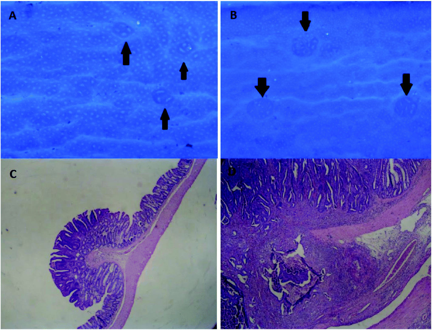
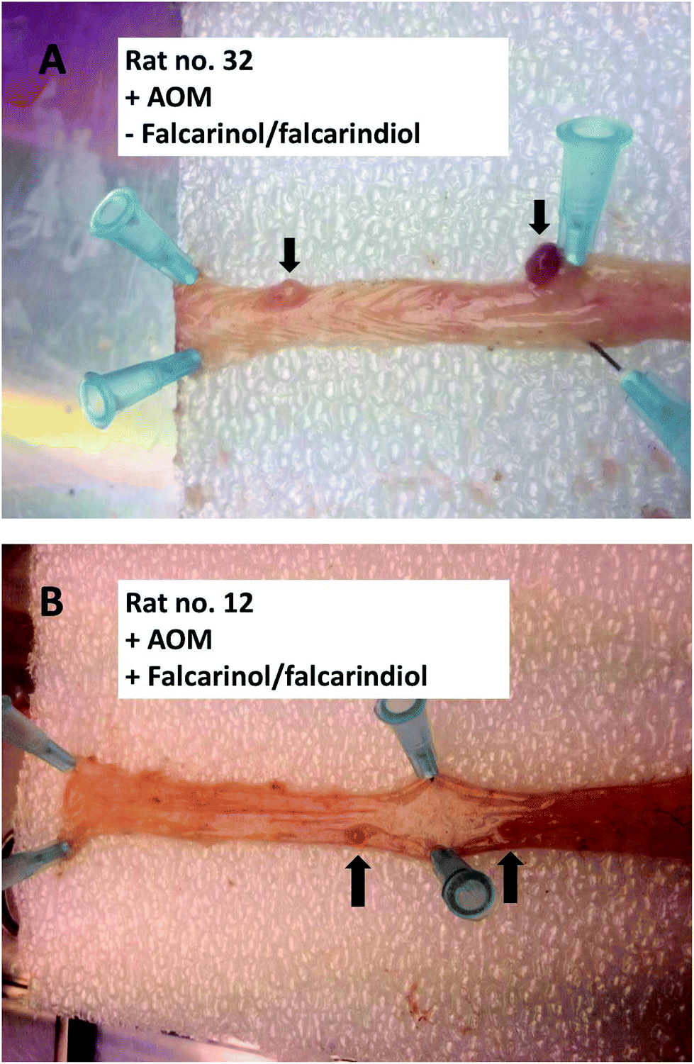
The rats were euthanized after 18 weeks and autopsied to examine for macroscopic alterations. The animals were killed by cervical dislocation, after they had been anaesthetized with isoflurane inhalation. Immediately after death, selected organs were fixed in 4% phosphate-buffered formaldehyde, pH 7.4, for later histopathological examination. The total length of the intestine was measured, and it was then cut longitudinally, rinsed in 0.9% NaCl solution, and pinned on a plastic slab. Before fixation, the large intestine was evaluated for macroscopic neoplasms, where diameter and location in the intestine were registered.
After fixation of the large intestine, Giemsa stain [6 mL
of stock solution (The Central Pharmacy at the Odense University
Hospital) in 50 mL of phosphate-buffered saline (PBS), pH 7.2, for 15
min] was used to visualize the ACF, and excess stain was rinsed off with
PBS. The tissue was placed with the luminal side up in a Petri dish
with enough PBS to cover the tissue. The total numbers of ACF and
tumours for each section were counted independently by two persons,
blinded to treatment modality, by using a stereomicroscope at 40×
magnification. The aberrant crypts were distinguished by their increased
size and thicker and deeply stained epithelial lining as compared with
normal crypts (Fig. 2).
An ACF may consist of one to several crypts, and in the present study,
the ACF were classified as small (1–7 crypts) or large (more than seven
crypts), while neoplasms 1–3 mm in diameter were classified as minor
tumours. Tumours more than 3 mm in diameters were classified as large
tumours. Within each class of lesions, the variation coefficient of the
two counts was less than 10%. The two counts were averaged.
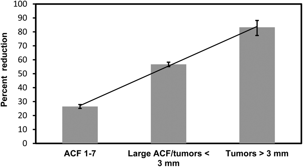
Macroscopic findings were fixed in 4% (v/v) formaldehyde buffered with 0.075 M sodium phosphate (pH 7) and embedded in paraffin. The tissues from tumours >3 mm were cut into 5 μm sections and were stained with hematoxylin and eosin. Additional sections were cut until characterization of the neoplasm was certain. The sum of lesions from large ACF and small macroscopic tumours were used in the calculation of the percentage reduction, because of the two categories were overlapping findings.
The occurrence of macroscopic tumours, and tumours larger than 3 mm, was tested between control and treated rats using a chi-square test. Data were analysed using exact methods for binomial data. A chi-squared test was used to assess the hypothesis of no difference in risk. Estimates are given with 95% confidence intervals. The number of ACF was tested between the groups using Wilcoxon rank sum test, data were analysed as two independent samples from the same distributions based on the Wilcoxon-signed rank. The assumption of equal distribution was checked by means and variances. Estimates are given with 95% confidence intervals.
FaOH and FaDOH were isolated from organic grown carrots of the cultivar Miami by flash column chromatography and preparative HPLC. The purified polyacetylenes (purity > 99%) were identified as (3R)-FaOH and (3R,8S)-FaDOH (Fig. 1) by spectrometric and spectroscopic techniques (see Materials and methods). Falcarinol type polyacetylenes like most polyacetylenes are thermally unstable and furthermore may undergo photodecomposition if exposed to UV light as well as oxidative and pH-dependent decomposition.13,17,41 However, regularly control of the purified polyacetylene standards stored in EtOH at −20 °C by analytical RP-HPLC and NMR did not show any sign of degradation, oxidation or isomerization of these compounds in the testing period of 19 weeks. The contents of FaOH and FaDOH were determined in all batches of feed of the active arm by LC-MS/MS, although a few batches from week 5–6, 7–8, 9–11 and 16–18 were combined, respectively, and analysed as one batch in triplicate (Table 1). The control rat diet contained no polyacetylenes (data not shown) and the mean contents of FaOH and FaDOH in the feed of the active arm were 6.88 ± 0.76 μg g−1 feed and 6.70 ± 0.77 μg g−1 feed, respectively (Table 1). Furthermore, analyses of random samplings from batches of feed of the active arm during the feeding experiments showed no sign of degradation, oxidation or isomerization of FaOH and FaDOH in accordance with the fact that no significant changes in the content of these polyacetylenes were observed (data not shown). It can therefore be concluded that the feed of the active arm contained the prescribed amount of FaOH and FaDOH of 7 μg FaOH per g feed and 7 μg FaDOH per g feed during the whole trial.
Fifteen out of twenty control rats and eight out of twenty FaOH/FaDOH treated rats had macroscopic tumours (P = 0.027) [Fig. 3]. No drop outs due to death or disease occurred before they were euthanized. All results are summarized in Table 2. The median number of small ACF was 218 in control and 145 in treated rats (P
< 0.001). The total numbers of neoplasia identified under
macroscopic examination of the rat intestine were 21 tumours in the
group of control rats and 12 in the FaOH/FaDOH treated rats. Number of
tumours larger than 3 mm were 6 in the group of control rats and 1 in
the FaOH/FaDOH treated rats (P = 0.032).
Tumours larger than 3 mm were confirmed as neoplastic by histological
analyses. No adenocarcinomas developed in the observation period.
Table 2 Total numbers of neoplastic lesions in rats receiving diet containing FaOH and FaDOH compared to rats receiving control diet
| Number of small (1–7) ACF clusters | Number of large (>7) ACF clusters | Minor tumors 1–3 mm | Number of tumors >3 mm | |
|---|---|---|---|---|
| Rats receiving control diet (n = 20) | 4094 | 126 | 15 | 6 |
| Median = 218 | ||||
| Rats receiving diet containing FaOH/FaDOH (n = 20) | 3007 | 50 | 11 | 1 |
| Median = 145 | ||||
| Percent reductiona | 26.6 (P < 0.001) | 56.7 (P = 0.027) | 83.3 (P = 0.032) | |
| a Percent reduction of lesions in each category is indicated with the significance level in parenthesis. Large ACF and small macroscopic tumours were overlapping findings; thus their sum was used in the calculation of the percentage reduction of these categories of neoplastic lesions. |
The number of lesions in the group of rats
receiving feed containing FaOH and FaDOH compared to the rats receiving
control feed, showed a linear correlation to size (Fig. 4).
The reduction in percentage for small ACF was 33.6%, and for large ACF
and small macroscopic tumours it was 56.7% and for tumours larger than 3
mm it was 83.3%. In addition, 11 neoplasms were found in the small
bowel of rats receiving feed containing FaOH and FaDOH and 17 neoplasms
in the rats receiving control feed.
| Fig. 4 Percent reduction of lesions in FaOH and FaDOH treated animals compared to control group. The reduction in the size of lesions was linear correlated (R2 > 0.9987). |
Colorectal cancer results from changes in cell transformation of colonic epithelial tissue with the initial development of dysplastic crypts, adenomatous polyps and then cancer.42–44 AOM is a potent carcinogen that has been shown to be an efficient inducer of colorectal cancer in rats,42,43 and it has been shown that the AOM-induced rat model represents a good predictor of chemopreventive efficacy in humans.45
In the early stages of colorectal carcinogenesis, dysplasia in the intestinal mucosa occurs with variable degrees of complexity, called aberrant crypt foci (ACF).42 In the AOM-induced rat model ACF are considered as putative preneoplastic lesions in the colon epithelium and ACF can be used as biomarkers to assess the efficacy of potential chemopreventive agents.46 Some of these lesions are precursors of adenomatous polyps and colorectal adenomas in rats and humans.47 However, only a proportion of the neoplastic lesions will develop into adenocarcinomas. The proportion of ACF that develop into macroscopic adenomas and further into cancer is unknown, but the pathway is generally accepted. The small ACF was significantly reduced by approximately 34% in the FaOH/FaDOH group compared to controls and for the large ACF (>7) a reduction of approximately 57% was observed, this reduction was also significant (Table 2). However, as large ACF and small macroscopic tumours were overlapping findings their sum were used in the calculation of the percentage reduction of these categories of neoplastic lesions (Table 2). In the group treated with FaOH/FaDOH the number of tumours larger than 3 mm was reduced by approximately 83% compared to the control group, and this difference was also significant (Table 2). Thus, it can be concluded that FaOH and FaDOH are able to inhibit neoplastic development in the colon. The effects observed in this study are, however, much more significant compared to those of our previous study,32 where an effect of carrots and FaOH (purity > 98%) treatments, respectively, was only observed on the development of tumours and large ACF, whereas no effect was observed on small ACF. In the present study the rat diet in the test group was supplemented with FaOH and FaDOH (purity > 99%) to the final concentration of 7 μg FaOH per g feed and 7 μg FaDOH per g feed thereby taken into account possible synergistic action of their antiproliferative activity.29 Furthermore, the concentrations of FaOH and FaDOH in this study are in total approximately 4-fold higher compared to the FaOH concentration used in our previous study. Altogether this may explain the significant inhibitory effect observed on all sizes of neoplastic lesions in the colon in the present study.
The intake of feed per day for a 200 g rat is approximately 20 g. Consequently, each rat in the test group ate approximately 0.14 mg FaOH and 0.14 mg FaDOH per day corresponding to 0.7 mg FaOH per kg and 0.7 mg FaDOH per kg rat per day. Due to the faster metabolic rate of rats compared to humans, metabolic calculations must be taken into account if one attempt to calculate equivalent doses to humans. A 70 kg person should therefore eat about 12 mg FaOH and 12 mg FaDOH daily to achieve a similar exposure if one assume that these polyacetylenes are readily available and are released in the colon. FaOH and FaDOH have been shown to be bioavailable and detectable in the systemic circulation in humans after oral intake of carrots,13,17 in accordance with their lipophilicity; hence, when released in the colon falcarinol type polyacetylenes possesses the ability to enter epithelial cells in the colon, although this requires further investigations.
The distribution of the most abundant polyacetylenes in carrots is known to vary among different genotypes. The content of these polyacetylenes in cultivated carrot cultivars varies from 8–424 mg per kg FW for FaDOH, 0.8–281 mg per kg FW for FaOH and 1–149 mg per kg FW for FaDOH3Ac.12,15 In addition the ratio between FaOH, FaDOH and FaDOH3Ac varies also with FaDOH often being the most abundant and FaDOH3Ac occurring in lowest concentration. The concentration of these polyacetylenes in the carrot cultivar D. carota ssp. sativus cv. Miami used for the isolation of FaOH and FaDOH was determined by analytical HPLC-PDA14 to be: 10.3 mg ± 0.9 mg FaOH per kg FW, 32.6 mg ± 3.1 mg FaDOH per kg FW and 3.6 mg ± 1.2 mg FaDOH3Ac per kg FW, and is in accordance with the concentrations and ratio of these polyacetylenes previously found in organic Danish grown carrot cultivars.12 Compared to cultured forms of carrots, the levels of FaOH and FaDOH in some D. carota wild relatives can be up to 10–20 times higher, whereas the differences in the content of FaDOH3Ac is less pronounced.15 The cultivated carrot cultivar ‘Nantes Empire’ has been reported to contain 46 mg FaOH per kg FW and 235 mg FaDOH per kg FW,15 which means that a person of 70 kg should eat around 260 g of this cultivar to obtain the effective dose of both polyacetylenes, if we assume that all the content of polyacetylenes in the fresh carrots is released in the colon. However, the wild relative D. carota ssp. carota has been reported to contain 1073 mg FaOH per kg FW and 1003 mg FaDOH per kg FW.15 This means that a person of 70 kg should only eat 12 g carrots per day to obtain the same exposure of FaOH and FaDOH as in the present preclinical trial. Consequently, it is realistic to assume that the amounts of FaOH and FaDOH used for testing in this preclinical trial can be achieved by a daily intake of for example fresh carrots or in the form of fresh carrot juice or smoothies. FaDOH3Ac may also contribute to the anticancer effect of carrots. However, as this polyacetylene has only been detected in carrots and usually occur in much lower concentrations compared to FaOH and FaDOH, as also demonstrated in the present investigation, its contribution to the health promoting effects of vegetables containing falcarinol type polyacetylenes is considered to be relatively limited. FaDOH3Ac was therefore not attempted isolated and included in this preclinical trial.
After subcutaneous injection of AOM in the rat, it is entering the circulation and the carcinogen is metabolized in the liver and then excreted to the intestinal system, resulting in macroscopic tumours of the colon and rectum, which was observed in 15 control rats out of 20 receiving AOM in the present study. The high incidence of small bowel tumours observed in the duodenum indicates that the carcinogens are excreted to the intestinal system via the bile system. Due to the fact that rats do not have a gall bladder, the highly reactive metabolites are excreted together with the bile directly into the duodenum of the rat; hence, the excretion of the carcinogens is not coordinated with feed intake. The carcinogens are mixed with the feed in the duodenum, and transported to the large intestine where they induce neoplasms in the colon of the rats. The mechanism by which the polyacetylenes prevent the neoplastic development is unknown; however, parts of the mechanism might be connected with direct interaction between FaOH and FaDOH and the carcinogen or metabolites from the carcinogen, thus preventing hypermethylation of the intestinal cells. The reduction in growth rate of the neoplastic lesions observed in the present study is, however, executed later, when AOM is no longer present in the rats, and therefore the observed inhibitory effect on the formation of neoplastic lesions of FaOH and FaDOH must be due to different modes of action. It has previously been shown that inhibition of COX-2 reduces the incidence of preneoplastic cells in rats treated with AOM.48,49 FaDOH has shown to be an effective inhibitor of COX-1 and COX-2, whereas the COX inhibitory activity of FaOH is less pronounced.17,21,33,50,51 In addition FaOH and FaDOH have shown to inhibit the formation of the pleiotropic proinflammatory cytokines interleukin-6 (IL-6), IL-1β and tumour necrosis factor (TNF)-α,16 and thus the activation of their upstream NF-κB, signalling pathway, which is crucial for neoplastic transformation and promotion.52 Accordingly FaOH and FaDOH are also strong inhibitors of LOXs, including 5-, 12- and 15-LOX,17,20 that are also involved in tumour-progression and activation of NF-κB. Although the COX inhibitory activity of FaDOH and FaOH might add to the explanation of the observed antineoplastic effect in this investigation, it may not be the only explanation as mentioned above and in the introduction; hence the mechanisms of action of the preventive effect of FaOH and FaDOH on colorectal cancer requires further investigations. It is, however, encouraging that the dose used in the present investigation of these polyacetylenes is within the range of normal daily intake of carrots, which does not cause any toxic effects in humans. FaOH has so far only shown unwanted toxic effects when delivered in high doses upon injection (100 mg kg−1) to rodents, where it causes neurotoxic symptoms,53 whereas FaDOH does not seem to have any toxic effect.54 Unwanted toxic effects of FaOH and FaDOH in humans by even a relatively high intake of carrots and/or processed carrot products or other Apiaceae vegetables is therefore not expected.
The present study has demonstrated that dietary supplements of FaOH and FaDOH reduced the number of neoplastic lesions formed as well as the growth rate of the polyps suggesting a preventive effect of these polyacetylenes on the development of colorectal cancer in AOM-induced rats. Furthermore, the amounts of polyacetylenes used in the preclinical trial can be achieved by a daily normal intake of carrots. Moreover it is not unlikely to assume that a normal intake of cultivated carrot cultivars containing high concentrations of FaOH and FaDOH will expose the colon for relative higher concentrations of polyacetylenes than in the present preclinical trial. Thus it could be interesting to determine the concentration of FaOH and FaDOH where an optimal preventive effect on neoplastic lesions in the colon is achieved in a FaOH/FaDOH dose–response preclinical trial. Eventually, such a study could be important for the preparation of clinical trials investigating the preventive effect of carrots and/or processed carrot products on the formation of neoplastic lesions and on the development of colorectal cancer in humans. It will also be obvious to investigate the possible mechanisms of action of falcarinol type polyacetylenes in preclinical and clinical trials, and in particular FaOH and FaDOH as they appear to be the most important dietary polyacetylenes based on the present and previous studies.
| ACF | Aberrant crypt foci |
| ACN | Acetonitrile |
| AOM | Azoxymethane |
| Caco-2 | Human epithelial colorectal adenocarcinoma cells |
| COX | Cyclooxygenase |
| EtOAc | Ethyl acetate |
| EtOH | Ethanol |
| FaOH | Falcarinol |
| FaDOH | Falcarindiol |
| FaDOH3Ac | Falcarindiol 3-acetate |
| FW | Fresh weight |
| IL | Interleukin |
| LOX | Lipoxygenase |
| NF-κB | Nuclear factor kappa-light-chain-enhancer of activated B cells |
| PBS | Phosphate-buffered saline |
| TFA | Trifluoroacetic acid |
The authors declare no conflict of interest.
We thank Dr Kai Grevsen Department of Food Science, Aarhus University, Denmark and the nursery Vostrup Øko, Denmark for the carrots. We also thank Department of Pathology at Odense University Hospital for technical support during evaluation of the study. Finally, we thank Biomedical Laboratory at University of Southern Denmark for technical support of the animal experiments.
Trust God, not sinful people. Worship God, but not things/people. Let God lead you, but not let others lead you. 相信上帝,而不是有罪的人。 敬拜神,而不是事/人。 讓上帝帶領你,但不要讓別人帶領你。
